Known Non-Nacreous Non-Bead Cultured Pearls and Similar Unknown Pearls of Likely Cultured Origin from Pinctada maxima
ABSTRACT
Non-bead cultured (NBC) pearls originating from the Pinctada maxima mollusk are commonly nacreous and white, cream, silver, or golden in color. This study details the characteristics of six non-nacreous NBC pearls and a partially non-nacreous NBC pearl recovered directly from Pinctada maxima mollusks during field trips to pearl farms in Australia and the results of their examination after applying microscopy, real-time microradiography (RTX), X-ray computed microtomography (μ-CT), long-wave ultraviolet (LWUV) radiation, DiamondView imaging, ultraviolet-visible (UV-Vis) spectroscopy, Raman and photoluminescence (PL) spectroscopy, and Fourier-transform infrared (FTIR) spectroscopy. An additional eight similar-looking pearls that were removed from a very large bag of reportedly NBC pearls were also examined using the same gemological and advanced identification techniques. Their results are also included and revealed features consistent with the six known NBC samples. While the internal structures of all the pearls are more characteristic of Pinctada maxima pearls, their external appearances could be confused with some other non-nacreous pearls such as those from the Pinnidae family (pen pearls), in particular. Raman and PL spectroscopy allow these pearls to be separated from pen pearls. Also included for comparison and discussion is a partially non-nacreous NBC pearl with an interesting internal structure that was sourced from a pearl farm by a contact in Australia and removed from the gonad of an operated mollusk.
INTRODUCTION
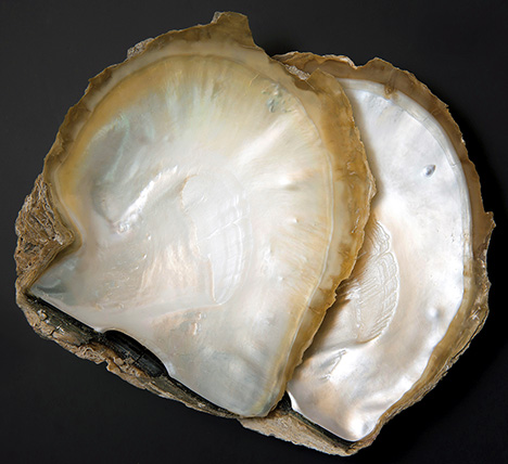
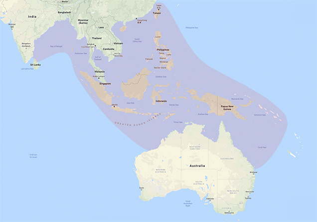
The Pinctada maxima mollusk, commonly referred to as the “South Sea pearl oyster,” is the largest of the Pinctada genus. Silver-lipped and gold-lipped forms of the mollusk, their names derived from the color of the nacreous rim on the inner mother-of-pearl (MOP) area of the shell (figure 1) as well as the underlying MOP surface beneath the periostracum, are commonly found. The outer surface of Pinctada maxima shells usually exhibits a light beige color with a series of ridges but lacks radial structure. In addition to the nacreous MOP surface on the inner part of the shell, a non-nacreous beige-colored lip consisting of clear prismatic crystalline ultrastructure is usually observed (Dauphin et al., 2019). The mollusk inhabits a wide area of the Pacific Ocean from the Bay of Bengal to the Solomon Islands and from Hainan, off the coast of China, down to the eastern and western coasts of Australia (figure 2) (Southgate and Lucas, 2008).
Wild and hatchery-bred Pinctada maxima shells are used on pearl farms mainly in the waters around Australia, Indonesia, the Philippines, and Myanmar. Bead cultured pearls produced by the mollusk range from 8 to 20 millimeters depending on the size of the host, the bead nucleus used, environmental conditions, and the health of the host. During the nucleation process, the mollusk is often chemically relaxed (Kishore et al., 2015; Zhu et al., 2019). Once the shell opens sufficiently, a skilled technician makes a small incision in the gonad in which a bead nucleus and piece of mantle tissue1 are inserted (Southgate and Lucas, 2008; GIA Pearls course, 2011). The mantle subsequently enables a pearl sac to develop around the nucleus, and the epithelial cells deposit nacreous layers for as long as conditions are suitable for the host to do so. If the mollusk ejects the bead, or the tissue and bead separate, the mantle tissue piece may nevertheless develop to a pearl sac and produce a non-bead cultured pearl, often referred to in the trade as a “keshi” pearl (Hänni, 2006).
While the vast majority of non-bead cultured pearls formed by Pinctada maxima mollusks are nacreous, this report will focus on lesser-known examples that are non-nacreous. Such pearls are more commonly associated with mollusks that do not belong to the Pteriidae family and may be divided into two distinct types: those that possess a flame-like pattern (also sometimes referred to as “porcelaneous”) produced by some mollusks (i.e., Lobatus spp., Melo spp., and some Tridacninae family members) and those that do not exhibit flame structures but possess cellular or other patterned structures (i.e., Pinnidae, Pectinariidae, and some Veneridae family members). However, this study proves that beige to brown non-nacreous pearls form within Pinctada spp. mollusks before the nacre has had a chance to be deposited (Hänni, 2012).
MATERIALS AND METHODS
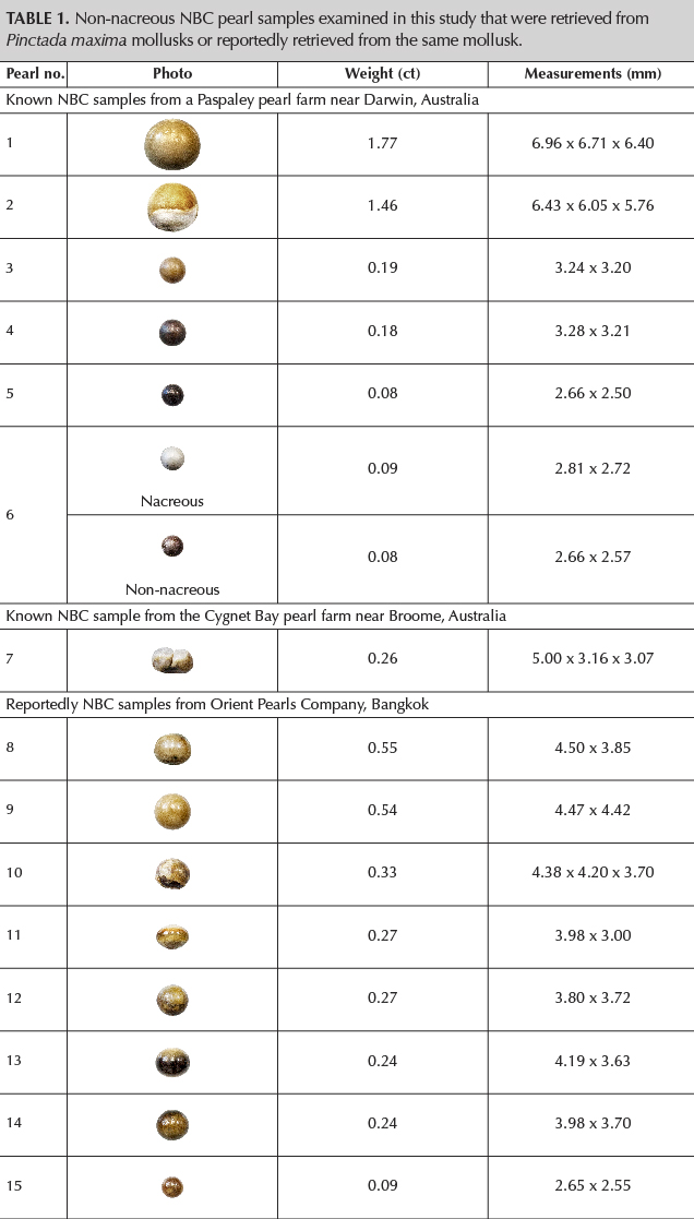
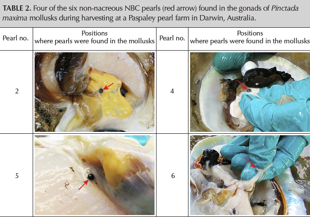
Six non-nacreous NBC pearls produced by Pinctada maxima mollusks (table 1, pearls 1–6) were recovered in October 2014 by GIA gemologists during a field trip to a pearl farm approximately 560 km southwest of Darwin, Australia, operated by Paspaley Pearls. When retrieved, five non-nacreous NBC pearls were found in the mollusks with bead cultured pearls and were recovered directly from the gonad. Another nacreous (when retrieved) NBC pearl (table 1, pearl 6 top) was found in the gonad area without a bead cultured pearl, but its nacreous layers separated from the underlying non-nacreous structure over time, leaving only the non-nacreous material showing later (table 1, pearl 6 bottom). The images captured when pearls 2 and 4–6 were retrieved from their shells are shown in table 2. One partially non-nacreous NBC pearl (table 1, pearl 7) was found in a Pinctada maxima mollusk by contacts of Ms. Laura Otter who own and run a pearl farm operated by Cygnet Bay Pearls (now Pearls of Australia), near Broome, Western Australia. In addition, eight likely NBC pearls with non-nacreous surfaces were loaned by Orient Pearls Bangkok (table 1, pearls 8–15). These eight pearls were removed from a large parcel of mostly nacreous pearls that were reportedly “cultured pearls” and were claimed to have been recovered from Pinctada maxima mollusks. The measurements of all the pearls ranged from 2.65 x 2.55 mm to 6.96 x 6.71 x 6.40 mm, and the weights ranged from 0.08 ct to 1.77 ct.
Real-time microradiography (RTX) and X-ray computed microtomography (μ-CT) analysis are the primary non-destructive techniques used to examine the internal structures of pearls. RTX images were obtained using a Pacific X-ray Imaging (PXI) GenX-90P X-ray system with a 4-micron microfocus, 90 kV voltage, and 0.18 mA current X-ray source, fitted with a PerkinElmer 4”/2” dual-view flat panel detector. μ-CT work was carried out with a ProCon CT-mini X-ray system with a 5-micron microfocus, 90 kV voltage, and a 0.18 mA X-ray current source. Two detectors were used to capture the results during this work: a Hamamatsu flat panel detector with 50 μm pixel pitch (pearls 1–6) and a Varex imaging flat panel detector with 74.8 μm pixel pitch (pearls 6–15), and a frame grabber card.
Photomicrographs of the surface structures were taken using a Nikon SMZ18 stereomicroscope with an SHR Plan Apo 1x objective lens and NIS-Elements imaging software.
The fluorescence reactions under LWUV radiation were observed under a GIA-designed LWUV unit incorporating a narrowband 365 nm UV LED source. A DTC DiamondView unit with a xenon flash lamp source and a special ultraviolet filter producing short-wave UV radiation of less than 225 nm was also applied.
Raman and PL spectra were obtained using a Renishaw inVia reflex micro-Raman spectrometer system with a 514 nm argon-ion laser and an 830 nm diode laser at 50 mW excitation. Raman spectra were collected with the 514 nm laser set at 5–10% power and the 830 nm laser at 0.1–10% power. Three accumulations with an exposure time of 10 seconds were collected for each laser. PL spectra were collected using the 514 nm laser with the power set at 1–10% and applying one accumulation with an exposure time of 15 seconds.
The UV-Vis spectra were collected in the 200–800 nm range with an Agilent Cary 60 UV-Vis spectrophotometer utilizing a custom-made GIA integrated sphere accessory incorporating an 80 Hz xenon flash lamp source.
FTIR reflectance spectra were recorded with a Thermo Scientific Nicolet iN10 infrared microscope using a 150 x 150 μm aperture. All data were collected with 100 scans per spectrum in the range of 675–7000 cm–1 using a liquid-nitrogen-cooled MCT-A detector system and a spectral resolution of 4 cm–1. A background spectrum was collected in air before each sample spectrum was obtained.
OBSERVATIONS AND RESULTS
External Appearances. The shapes of the majority of pearls varied from round and near-round to button and oval. The colors ranged from cream to black, and most of them exhibited more than one color or differed in saturation and tone over different areas of the same pearl.
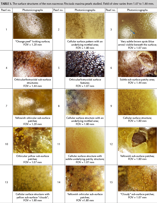
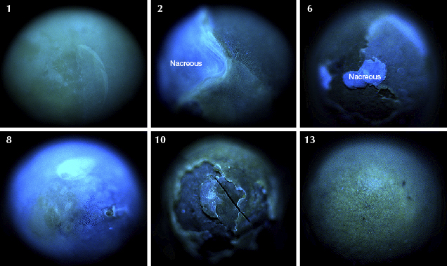
Through the microscope, all but two of the pearls lacked the clear overlapping platelet structure typically associated with nacreous pearls (table 3). Only pearls 2 and 7 exhibited nacre, but only on one white area of each pearl. The majority of pearl 2’s surface resembled the others and displayed a non-nacreous structure, evident as a cellular pattern on a light brown area, and a patchier region below the surface in a cream-colored area. Cellular structures of differing sizes and shapes were evident on the non-nacreous part of pearl 2 and over the entire surface area of pearls 8, 9, 11, and 13. The surface condition of most pearls was poor, as they showed rough and dull features. Most of the pearls only showed a patchy appearance that lacked clear structure on or below their surfaces. Yellowish patches were present below the surfaces of non-nacreous areas of pearl 7 and pearls 10, 12, and 14, while minute brown features (table 3, blue arrow) were present below the surfaces of pearls 3, 11, and 14. Orbicular/botryoidal features were present all over the surface of pearl 5.
Internal Structures. RTX and μ-CT analysis was performed on all the pearls (tables 4 and 5). None of the pearls revealed a shell bead nucleus, which is not surprising as non-nacreous pearls of a similar appearance are not commercially available and have only rarely been seen in the market (Segura and Fritsch, 2014).
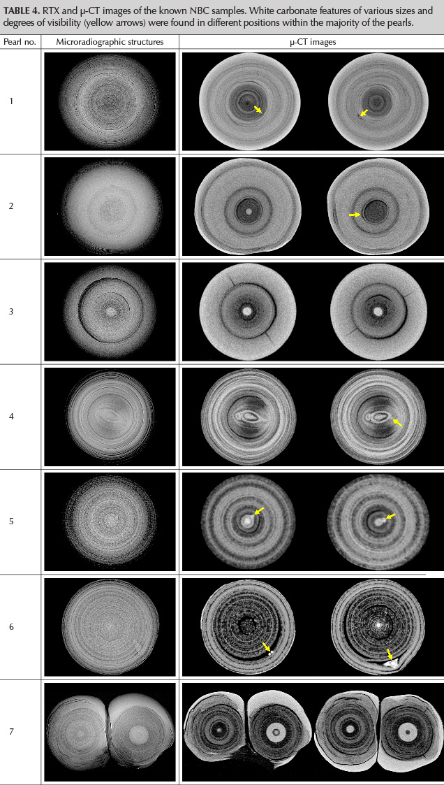
RTX analysis of the known samples revealed cores of different sizes and forms—dark or white— the majority surrounded by more radio-translucent organic-rich structures that show as darker areas owing to the easier passage of the X-rays. The more radio-opaque areas show as lighter areas where the X-rays do not pass through so readily. The cores varied from tiny to very large, mostly round ones, and the shape of the core in pearl 4 clearly appeared “off-round” (pear-shaped). The mostly organic-rich concentric layers around the cores showed a variety of growth stages, with rings that were markedly clearer and more defined than other stages in the pearls’ formation. All these growth patterns may be seen in natural and NBC pearls (Nilpetploy et al., 2018b). An uneven or “spotty” pattern was visible in some of the pearls, and radial structure was present in pearls 1–3. While these structures are questionable when combined and are not typical natural structures, they still do not offer conclusive proof of NBC pearls. μ-CT analysis provided additional structural features and helped clarify the RTX results. White carbonate (Krzemnicki et al., 2010, 2011) features (yellow arrows in tables 4) of various sizes and shapes were found in different positions with the majority of the pearls. Only pearl 3 did not show these features. A small void was visible in one feature next to the open ovoid “core” of pearl 4. The clearer details provided by μ-CT analysis revealed features that were too suspicious to characterize some of the pearls as “natural” non-nacreous pearls and in the authors’ opinions provided sufficient evidence to classify them as NBC pearls. This is especially true of pearls 4–6 from the group that were found in the gonads of Pinctada maxima mollusks. Pearl 4’s unusually shaped and more complex core structure together with a very small white “seed” feature in the core area is more typical of NBC pearls examined by GIA in previous research (Nilpetploy et al., 2018a). Very similar seed features are evident in pearls 5 and 6 to varying degrees as well. However, pearls 1–3 could readily pass as natural pearls if their gonad origin was not known, and this highlights the challenges of accurately identifying all pearls based only on their structure. It is inevitable that some NBC pearls are called natural and some natural are labeled as NBC by laboratories when their full history is unknown during examination, which is the case with the vast majority of pearls that pass through gemological laboratories today.
Both parts of pearl 7 revealed cores of different sizes and structures surrounded by more radio-translucent organic-rich areas. While the large core in the right-hand section appears suspect, it also exhibits a smaller dark organic core at its center. No obvious seed features were observed, but a “spotty” pattern present in the organic-rich areas is similar to some of the internal features noted in the known NBC pearls from a Paspaley pearl farm. Although the overall structure is quite similar to that expected for natural pearls (Nilpetploy et al., 2018b), the spotty nature of the organic-rich areas and large white core within one half of the twin would be enough to generate some concern about its identity. Yet, even though it is a known NBC pearl recovered from the gonad of a mollusk, it would be very challenging in laboratory conditions to issue a report stating it is an NBC pearl.
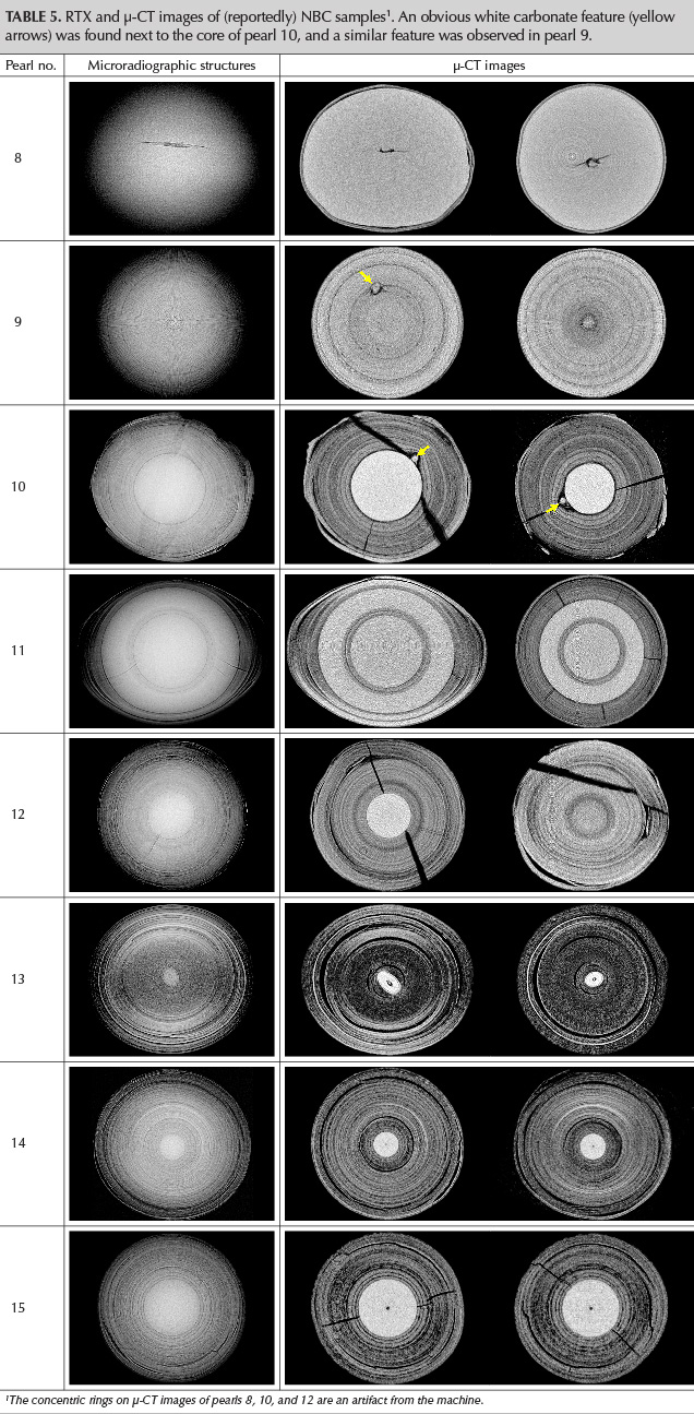
Although pearls 8–15 (table 5) were not personally recovered from the gonads of Pinctada maxima mollusks but were instead mixed with claimed nacreous NBC pearls from Pinctada maxima mollusks in one large bag, it is striking to see the similarities their internal features share with those in table 4 that were retrieved from the gonads. Only pearl 8 shows a suspect linear-void feature that is often associated with NBC pearls (Hänni, 2006; Sturman, 2009a; Sturman et al., 2016; Nilpetploy et al., 2018a; Manustrong, 2018). RTX analysis of pearl 9 revealed a distinct radial structure similar to that seen in natural non-nacreous pen pearls (Sturman et al., 2014), but a suspect irregular feature was clearly visible within the growth layers when the μ-CT data was reviewed. Although it is not a definite NBC pearl, the odd feature within the concentric and radial body is enough, in our opinion, to bring its identity into question. Seed features are present in pearls 9 and 10 (yellow arrows in table 5), while pearls 10–12 and 15 show large to very large white cores. Pearl 15 shows a very small dark feature at the center of the white core, and its overall structure is suspicious enough to raise some red flags. The ovoid shape of the core in pearl 13 also generates sufficient concern about its identity and would fit more with an NBC pearl than a natural one. Only its non-nacreous external appearance would lead to doubts about its NBC identity.
External Composition. All the pearls were examined using a Raman spectrometer in order to obtain their Raman and PL spectra. Raman spectra collected using a 514 nm laser (figure 3) revealed very weak peaks at 1086 cm–1 associated with aragonite and calcite, but no other clear peaks were detected to identify which of the two calcium carbonate polymorphs were present in the pearls (Urmos et al., 1991).

Since the spectra obtained using the 514 nm laser failed to clearly identify which calcium carbonate polymorphs were present, all of them were also examined using an 830 nm laser (figure 4). All the pearls revealed a more discernable peak at 1085 cm–1. More importantly, pearls 1–3, 6–8, 10–13, and 15 revealed doublet peaks related to the vibration modes of aragonite crystals at 702 and 705 cm–1. The more lustrous non-nacreous area on pearl 2 and some areas on pearl 9 revealed a peak at 713 cm–1 and another at 280 cm–1 known to be characteristic of calcite. The use of a different laser therefore permitted us to identify the calcium carbonate polymorph that formed the external surface layers of most samples. Only pearls 4, 5, and 14 still failed to show any clear features.

The PL spectra obtained were similar in form to one another with the maxima centered around 610 nm. The wavy pattern observed in the 550–700 nm range, possibly resulting from the non-nacreous structure, was consistent in all the pearls (figure 5). As would be expected from some of the Raman results, 12 of the pearls exhibited marked fluorescence reactions, as can be seen at higher wavelengths.

Even though the 830 nm laser proved which calcium carbonate polymorphs were present on 11 pearls, three pearls still failed to show any clear features. This was better than the results obtained using the Raman microscope’s 514 nm laser, which proved inconclusive for all the samples. Hence, all of them were also examined using an FTIR spectrometer. The resulting FTIR spectra revealed peaks related to the vibration modes of an aragonite crystal lattice at 700 (v4b), 713 (v4a), 877 (v2), 1083 (v1), 1440 (v3b), and 1473 (v3a) cm–1 (figure 6) (Adler and Kerr, 1962; Andersen and Brečević, 1991; Ramasamy et al., 2003; Tan et al., 2005; Wang et al., 2006; Otter et al., 2017). Although the more lustrous non-nacreous area on pearl 2 and the cellular area on pearl 9 revealed peaks at 889 and 1440 cm–1 (figure 7), the absence of peaks at 700 and 1083 cm–1 and the presence of a peak at 1440 cm–1 when associated with a 711 cm–1 peak are calcite related. The FTIR and 830 nm Raman laser results seem to correspond and confirm that the surface compositions on areas of pearls 2 and 9 differ.


UV-Vis Spectrophotometry. UV-Vis reflectance spectra were obtained for all the pearls apart from those below 3.20 mm. The latter were too small for the aperture of the diffuse reflectance accessory used and as a result could potentially damage the accessory’s integrated sphere. The resulting spectra showed similar patterns in the visible region, with a slope from the red region at 750 nm where the maxima occurred to minima at 400 nm, as expected for dark brown to black pearls not originating from mollusks of the Pteria species or nacreous pearls from Pinctada margaritifera (Nilpetploy et al., 2018a), Pinctada mazatlanica (Homkrajae, 2016), or Pinctada maculata (Nilpetploy et al., 2018b). The darker-colored pearls exhibited a lower reflectance than the lighter-colored pearls, as can be seen from the spectra of pearls 8 and 13, which show spectra with higher reflectance that correlates to their lighter bodycolors (figure 8). While a few of the pearls showed a weak 280 nm feature, it was most evident in pearl 8, which has the lightest color in the group. A small area with higher fluorescence is also visible in the spectrum of pearl 8 in particular, at approximately 310 nm. It is much weaker in some of the other pearls and absent in the darkest. This feature has been observed in many Pinctada species pearls (Elen, 2002; Karampelas et al., 2011; Karampelas, 2012; Nilpetploy et al., 2018a and b), some Lobatus gigas2 (Queen conch) pearls, Mercenaria (quahog) pearls, and freshwater pearls. The 310 nm feature has not been observed in Pinnidae family pearls (Sturman et al., 2014).
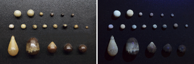
Luminescence. Under LWUV radiation (figure 9), all the pearls exhibited nearly inert (very weak to weak yellowish green) to moderate-strong yellowish green and greenish blue fluorescence of various intensities. Some of the pearls exhibited an uneven pattern, in keeping with their inhomogeneous coloration (figure 9, top and middle rows). The nacreous portion of pearl 2 exhibited moderate greenish yellow fluorescence, which differed markedly from the other samples. The nacreous portion of pearl 7 exhibited weak yellowish green fluorescence, and organic-rich areas were inert to weak yellow. When compared to pearls that reportedly originated from Pinnidae family (pen pearls) mollusks (Sturman et al., 2014), these non-nacreous Pinctada maxima samples exhibited more of a bluish to green reaction than the yellowish reaction of some of the pen pearls (figure 9, bottom row). Their reactions also differed from the strong orangy red fluorescence often observed in non-nacreous Pteria species pearls described previously by the authors and others (Kiefert et al., 2004; Sturman, 2009b; Karampelas and Abdulla, 2017).
Reactions under the ultra-short UV wavelength radiation of the DiamondView were also observed on six of the pearls at random (figure 10). Their fluorescence reactions ranged from almost inert to a strong chalky blue with patchy yellow-green areas on some. The nacreous portion of pearl 2 exhibited a bright chalky blue fluorescence, in keeping with other nacreous Pinctada species examined previously (Scarratt et al., 2012), while the cellular structure exhibited a much weaker blue fluorescence with yellow-green boundaries and other areas of weak patchy yellow-green fluorescence. Two small nacreous areas on the surface of mostly non-nacreous pearl 6 also showed a moderate/strong chalky blue fluorescence while the rest of the pearl revealed an almost inert reaction.
CONCLUSION
While Pinctada maxima mollusks are mainly known for producing nacreous white, silver, and “golden” pearls, they may also produce non-nacreous pearls. In contrast to the nacreous pearls, non-nacreous pearls from Pinctada maxima are rare, and this study on a limited sample base shows they appear to form in sizes under 7 millimeters. The samples obtained for this study indicate that this type of material forms in quite symmetrical shapes. Irregular examples were not readily available, and only pearl 7 fell into this category. Most of the pearls examined in this work revealed uneven coloration, and their surface quality was poor owing to numerous cracks and “peeling.” Where cellular structure was present, the outline of each cell was more irregular than those in pen pearls, which are often more elongated (Sturman et al., 2014), and also differed from the non-nacreous areas seen on Pteria species pearls, which often appear more hexagonal (Karampelas et al., 2017). Although their yellowish green to greenish blue fluorescence under LWUV radiation looked quite similar to that shown by some lighter-toned pen pearls, the latter exhibited a slightly more yellow hue in their fluorescence. However, their LWUV fluorescence clearly differentiates them from non-nacreous Pteria species pearls, which usually show a distinct orange to orangy red fluorescence.
Despite the similarities in the fluorescence reactions of the non-nacreous Pinctada maxima pearls with those of pen pearls, the differences in their Raman spectra readily aid in their separation. While the Pinctada maxima pearls show aragonite features around 702, 705, and 1086 cm–1, pen pearls generally revealed a clear calcite peak at ~712 cm–1 and another diagnostic calcite peak at ~280 cm–1. The PL spectra were also different from those seen in pen pearls, since the clear calcite peaks seen in pen pearls were absent. FTIR spectra revealed clear aragonite features on all the pearls except samples 2 and 9, which revealed obvious calcite features on some areas after analysis with the Raman’s 830 nm laser and subsequent FTIR work.
The UV-Vis reflectance spectra of the non-nacreous Pinctada maxima pearls did not reveal any obvious features. A weak one at approximately 280 nm was only present in 3 of the 11 pearls, but this feature was clear and consistent in the majority of pen pearls. The Pinctada maxima samples also failed to show either the 495 nm feature commonly observed in Pteria species pearls (Iwahashi and Akamatsu, 1994) or the same feature in combination with the 700 nm feature routinely detected in the spectra of nacreous Pinctada margaritifera and Pinctada mazatlanica pearls (Karampelas et al., 2011; Homkrajae, 2016; Nilpetploy et al., 2018a).
Although spectroscopy was able to separate the non-nacreous Pinctada maxima pearls from pen pearls and Pteria species pearls, the spectra did not show any diagnostic features to prove they were sourced from the Pinctada maxima mollusk. However, their defined concentric, sometimes radial and organic-rich internal structures are very characteristic of some pearls produced by the mollusk (Scarratt et al., 2012) and differ enough from other natural non-nacreous pearls to assist in the identification with a high degree of confidence. The structures are also, on the whole, suspicious enough to identify the pearls as being NBC rather than natural, although in some cases a difference of opinion would no doubt arise. It is also worth noting that attempts to find similar non-nacreous NBC pearls from akoya oysters inspected on a farm operated by Broken Bay Pearls Pty Ltd failed to yield any samples (Otter, 2017). This might have been because the farmer was not looking for such pearls or because they were markedly smaller than the samples obtained from their larger Pinctada maxima relatives and hence evaded observation completely. Some non-nacreous bead cultured akoya pearls recently examined by the authors from GIA Bangkok measured between 2.8 and 5.0 mm., but they were not NBC per the subject matter of this work and hence are not included.
FOOTNOTES |
|
↩ Back to article ↩ Back to article |


