Tubular Feature in a Natural Pearl
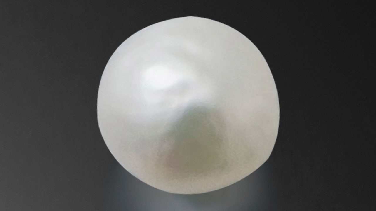
Recently, GIA’s Bangkok laboratory received a 1.27 ct white semi-baroque pearl for pearl identification service (figure 1). Under 40× magnification, the surface exhibited typical nacreous overlapping aragonite platelets (platy structure).

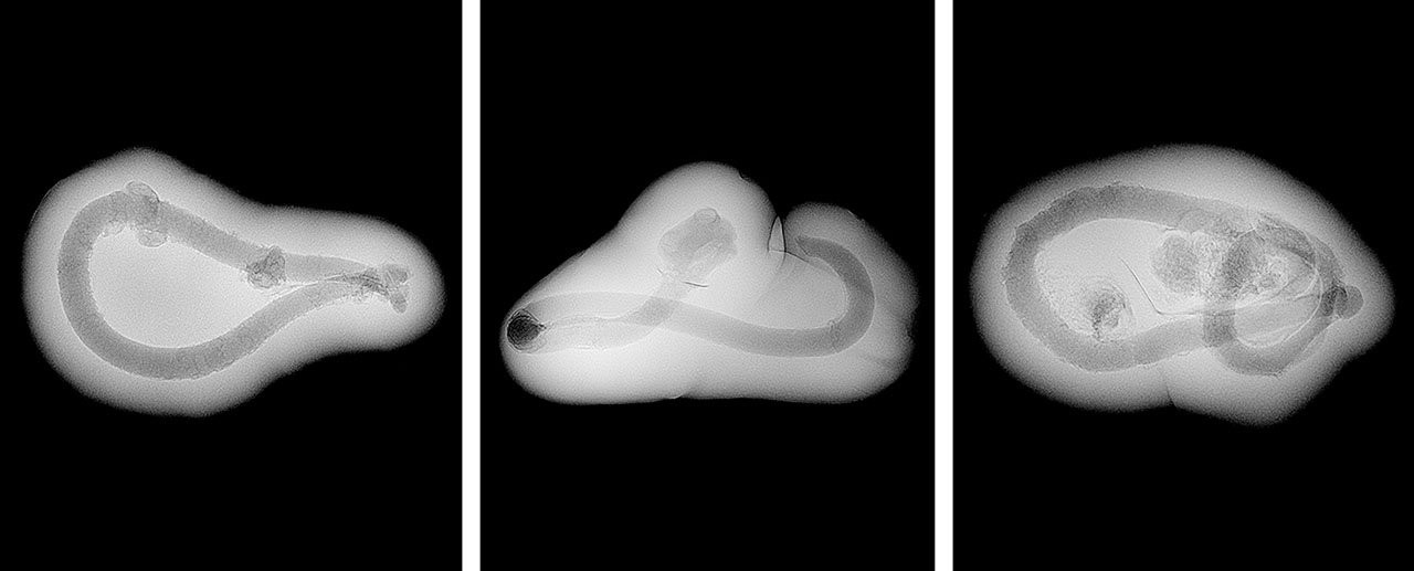
Real-time microradiography (RTX) revealed a tubular feature like a twisted tunnel at the center of the pearl, surrounded by fine growth arcs. When observed along the pearl’s thickest direction, the feature resembled a void or cavity-like structure (figure 2, left), similar to those found in non-bead cultured pearls (“The microradiographic structures of non-bead cultured pearls,” GIA Research News, November 20, 2009). However, when viewed from its flat direction (figure 2, center), the elongated tubular nature of the feature was inconsistent with the voids observed in non-bead cultured pearls. This inconsistency was further supported by X-ray computed microtomography (μ-CT), which showed multiple interconnected rounded voids (figure 2, right). When exposed to X-ray fluorescence, the pearl displayed an inert reaction indicative of a saltwater environment.
While this was the first time the lab had encountered a client submission with such a structure, similar structures were previously documented in three reference pearl samples from GIA’s research database. The three were reported to be natural Pinctada radiata pearls sourced from the waters of Bahrain; they were baroque and weighed 0.25, 0.34, and 0.44 ct (figure 3). Those samples showed a similar elongated twisted tubular feature at their center in RTX and μ-CT scan imaging, which helped confirm the natural origin of the examined pearl.
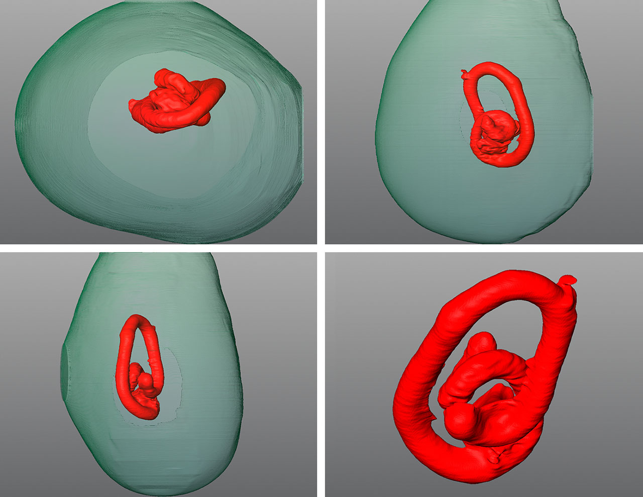
To further study this tubular feature, three-dimensional models were created using specialized software to render the μ-CT scan images (“New 3-D software expands GIA’s pearl identification capabilities,” GIA Research News, May 13, 2016). The results from these models clearly showed that the central structure consisted of an elongated twisted tube. Based on the tubular formation presented, the feature is suspected to be formed by a burrowing parasite trapped in the center of the pearl (figure 4; see video below). Parasites of various types have been observed in both saltwater and freshwater bivalves, and certain worms have been known to use bivalve mollusks as their hosts. Notably, marine bivalve mollusks tend to be more prone to parasitic intrusions than freshwater mussels (E. Strack, Pearls, Rühle-Diebener-Verlag, Stuttgart, 2006, p. 116). Therefore, the presence of such tubular features in saltwater pearls indicates the potential infiltration of worm larvae into the mollusks’ mantle. These larvae—either dead, alive, or hibernating—may have subsequently become encapsulated by the mantle epithelial cells, leading to the formation of a pearl sac and eventually the creation of a pearl.


