Unusual Large Nacreous Pen Pearls

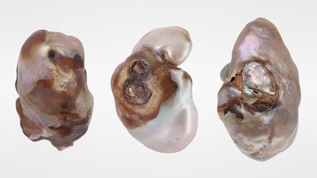
GIA’s Mumbai laboratory has received a wide variety of interesting pearls for identification since opening its pearl testing department in May 2022. One recent submission included three baroque pearls weighing 24.47 ct, 36.58 ct, and 36.70 ct and measuring 28.22 × 18.04 × 9.84 mm, 28.79 × 19.48 × 10.40 mm, and 32.49 × 20.50 × 18.35 mm, respectively (figure 1).

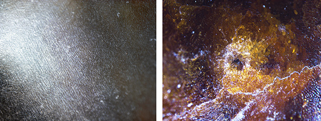
Visual observation revealed a combination of nacreous and non-nacreous surface structures. The pearls were white to light gray and brown and exhibited a strong orient with variations in saturation and tone. In some areas, their surface quality was poor to slightly damaged. Viewed under 40× magnification, the white to light gray areas displayed a striated and much finer form of nacre growth structure than observed in most nacreous pearls from Pinctada and Pteria mollusks (figure 2, left). The dark brown areas showed a characteristic non-nacreous cellular structure consisting of a network of closely packed cells (figure 2, right). These observations were consistent with previous studies (N. Sturman et al., “Observations on pearls reportedly from the Pinnidae family (pen pearls),” Fall 2014 G&G, pp. 202–215).
Raman analysis using a 514 nm and 830 nm laser excitation was performed on different surface areas. The nacreous areas on all three pearls showed a doublet at 701 and 704 cm–1 and a peak at 1085 cm–1 indicative of aragonite. However, the brown non-nacreous areas on pearls 1 and 2 showed a peak at 712 cm–1 indicative of calcite. Photoluminescence analysis for all pearls revealed weak bands at 620, 650, and 680 nm, suggesting natural coloration (S. Karampelas et al., “Raman spectroscopy of natural and cultured pearls and pearl producing mollusc shells,” Journal of Raman Spectroscopy, Vol. 51, No. 9, 2020, pp. 1–9).

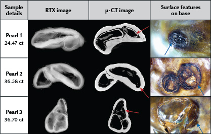
Despite the possibility of these three being large blister pearls, closer examination of them did not reveal any indications of previous attachment to the shell such as signs of heavily worked or sawn areas (“Natural shell blisters and blister pearls: What’s the difference?” GIA Research News, August 26, 2019). The lack of such features indicated they were whole pearls. The few small organic-rich areas visible on the pearls’ surfaces (figure 3), along with the non-nacreous and nacreous surfaces, were possibly damaged because the organic-rich material weakened over time.
X-ray fluorescence analysis revealed an inert reaction for all the samples. Energy-dispersive X-ray fluorescence spectrometry showed very low manganese levels (below detection limit in both pearls 1 and 2 and 9.9 ppm in pearl 3) and high strontium levels (1535 ppm, 1682 ppm, and 1250 ppm, respectively). Both analytical results were indicative of a saltwater origin.
Real-time microradiography (RTX) and X-ray computed microtomography (μ-CT) revealed a combination of large voids with some dark organic-rich material surrounded by white walls and fine growth arcs (figure 3). A chambered effect was notable in all three samples. The complex appearance of the voids differed from those observed in most non-bead cultured pearls from the Pinctada species, yet they were similar to nacreous and non-nacreous pen pearls reported in Sturman et al. (2014). The outline of the voids was consistent and followed the shape of the pearls’ surfaces, unlike the irregular and inconsistent voids found in the non-bead cultured pearls. In addition, μ-CT imaging revealed light gray and dark gray areas associated with the presence of organic matter within the voids, a characteristic commonly observed in natural pearls (“Non-bead cultured pearls from Pinctada margaritifera,” GIA Research News, April 27, 2018).
Based on the external and internal structure analysis, the samples were identified as natural pearls from the Pinnidae family (pen pearls). Visual observation, combined with the absence of any evidence of commercial culturing, played a crucial role in their identification. Their size and appearance further supported their classification as natural whole pearls.


