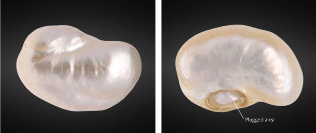
A Partially Hollow Natural Blister Pearl Filled with Foreign Materials

Over the centuries, India has maintained its reputation as a natural pearl trading center. Since the launch of GIA’s pearl identification service in Mumbai in May 2022, the laboratory has received a variety of interesting items for identification. One that stood out owing to its size and internal structure was a unique partially hollow natural blister pearl with unidentified fillers (figure 1).

The very large baroque white pearl weighed 169.05 ct (33.81 g) and measured 43.12 × 28.83 × 19.26 mm. However, on initial examination, its “heft” (perceived weight compared to its actual weight in comparison to its size) felt heavier than it should have. While the pearl’s face-up side exhibited minor surface blemishes and good nacre condition (figure 2A), its base showed a deep oval indentation that looked a bit suspect to the unaided eye and needed further detailed examination. Under 40× magnification, the area revealed a distinct boundary with remnants of a transparent adhesive around it (figure 2B). Evidence of heavy working and polishing on and around the base was also clearly visible (figure 2, C and D).
Many loose nacreous baroque or semi-baroque pearls of this size are hollow to some degree, and most turn out to be blister pearls that were attached to the shell and removed at some point (“An unusual pearl,” GIA Research News, March 31, 2009) or were shell blisters. In order to ensure the durability of these hollow pearls, and sometimes to add weight and increase their perceived value, they are filled with various materials such as metals, resins, waxes, shell, and other pearls (Fall 2013 Lab Notes, pp. 172–173) and fashioned to improve their external appearance. Although the initial examination indicated that this pearl was likely filled with foreign materials, further analysis was required.

Real-time microradiography revealed a large organic void/cavity filled with a radiopaque white material, probably small metal fragments or metal shavings (figure 3; see also Summer 2019 Lab Notes, pp. 251–254). Areas of greater radio-transparency suggested that some type of bonding or stabilizing material, possibly adhesive, may have been used to hold the foreign pieces in place, and there also appeared to be one other type of radiolucent material within the cavity. The pearl was examined in several directions in order to further analyze the internal structure and fillings. The outline of the void was smooth and blended with the external shape of the pearl. The outer nacre layers revealed fine growth arcs and a small organic-rich area was visible at the base, both of which aligned more with the growth features of a natural pearl. It was clear that this was originally a blister pearl or shell blister that had been cut from a shell, and that the indented area, which was the opening on the base of the pearl, was the point of attachment that was subsequently plugged. X-ray fluorescence proved that the pearl formed in a saltwater environment since most of its surface appeared inert and only a small area at the base displayed a very light greenish yellow reaction uncharacteristic of a freshwater environment.
Energy-dispersive X-ray fluorescence spectrometry on two areas (face and base) showed low or below detection levels of manganese and higher strontium levels, above 1000 ppm, both characteristic of a saltwater environment. The higher strontium levels coupled with the large size indicated that the mollusk from which the pearl originated was Pinctada maxima.
Raman analysis using a 514 nm laser excitation on two spots (face and base) of the surface produced high fluorescence due to the pearl’s light color and showed a doublet at 702 and 705 cm–1 as well as a peak at 1085 cm–1 indicative of aragonite. The photoluminescence spectra also displayed high fluorescence together with the aragonite peaks, as observed via Raman, typical of most nacreous pearls. Ultraviolet/visible/near-infrared spectra within the 220–850 nm range collected from the face and base were as expected for white to light cream-colored pearls and showed a predominantly high reflectance. The presence of a prominent 280 nm absorption feature coupled with moderate yellow reaction under long-wave UV excitation indicated that bleaching had most likely not been carried out.
After conducting a full analysis, GIA concluded that this sample most likely formed as a hollow natural blister pearl attached to its shell host. At some point, it was removed and the attachment area left an opening into the pearl. It was eventually worked and filled with foreign fillers (metal shavings/pieces and possibly some other materials) to improve its durability and make it suitable for setting; without the filling, it would not be usable in jewelry. The size, spectral characteristics, and chemistry were also consistent with a Pinctada maxima host mollusk.


