A Closer Look at Internal Etch Channels in Diamond

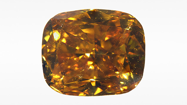
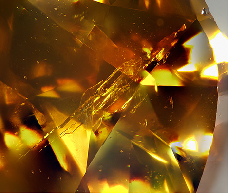
The Carlsbad laboratory recently received a 1.02 ct cushion-cut Fancy Deep brownish yellowish orange diamond (figure 1). The yellowish color of this type Ib/IaA diamond is mainly attributed to single substitutional nitrogen atoms in the diamond lattice. In addition to the presence of multiple pinpoint inclusions, the diamond contained a bundle of internal etch channels extending from a single irregular-shaped opening on the girdle (figures 1 and 2).


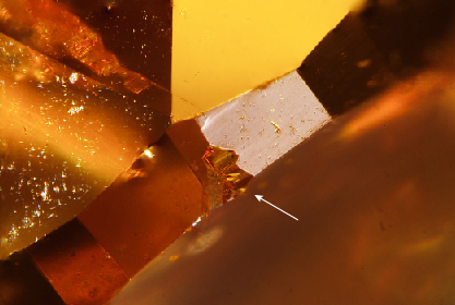
Narrow etch channels are rarely observed in gem diamonds. Etch channels can form due to dissolution of the diamond crystal by fluids/melts in the mantle, or during eruption to the surface (T. Lu et al., “Observation of etch channels in several natural diamonds,” Diamond and Related Materials, Vol. 10, No. 1, 2001, pp. 68–75; J.W. Harris et al., “Morphology of monocrystalline diamond and its inclusions,” Reviews in Mineralogy and Geochemistry, Vol. 88, No. 1, 2022, pp. 119–166). Defects in the diamond structure such as lattice dislocations are local areas of structural weakness, which are more susceptible to etching. In this diamond, multiple narrow etch channels originate from a single surface opening (figure 3), reflecting selective diamond dissolution as fluids permeated through the stone.

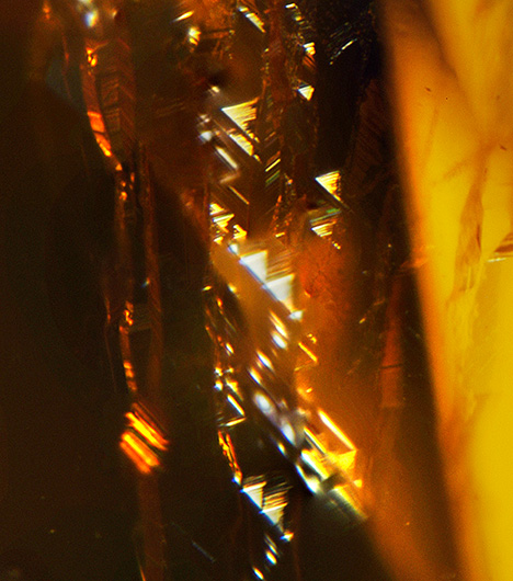
An intriguing feature observed along the walls of the internal etch channels consisted of multiple triangular etch pits known as trigons (figure 4). Trigons are one of the most common features found on the surfaces of rough diamonds, with various sizes, depths, and shapes (e.g., flat- or point-bottomed, attributed to etching by oxidizing fluids/melts; Harris et al., 2022). When present on diamond surfaces, trigons are restricted to the octahedral crystal face and may occasionally form parallel rows that follow the orientation of plastic deformation lines (often called “grain lines” in the trade, caused by dislocation of carbon atoms along octahedral planes). In faceted diamonds, trigons are usually removed by polishing but are occasionally preserved on the girdle. Conversely, trigons reported within internal diamond etch channels are extremely rare (Lu et al., 2001). The occurrence of trigons within a subsurface channel indicates etching along the octahedral crystal face of this diamond.


