Red-Dyed Spodumene Imitating Ruby
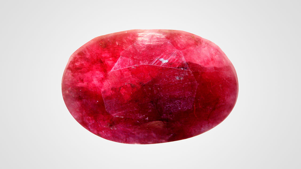
A semitranslucent deep red oval faceted gem, measuring 13.88 × 9.08 × 5.88 mm and weighing 5.72 ct (figure 1), was recently submitted to Stuller Inc. by a client for a custom design. The stone was presented as “red corundum.” But initial quality control testing showed a biaxial refractive index of 1.659–1.678 with a birefringence of 0.019, which did not correspond with corundum.
Due to the incorrect identity, the stone was sent to Stuller’s gemological laboratory for further analysis. Standard gemological testing revealed a specific gravity of 3.237, with a moderate orange reaction to long-wave UV and a very weak reaction, along the fractures, to short-wave UV, typical of spodumene species. Further analysis, using a 532 nm Raman laser, also provided well-matched results for spodumene (LiAl(SiO3)2). Although the collected results clearly pointed to spodumene, the deep red color it displayed was questionable and required further investigation.

Unpolarized ultraviolet/visible/near-infrared (UV-Vis-NIR) spectroscopy showed two merging broad bands centered at 530 and 561 nm (figure 2). The former is probably associated with the Mn3+ chromophore (H.U. Rehman et al., “An X-ray absorption near-edge structure (XANES) study on the oxidation state of chromophores in natural kunzite samples from Nuristan, Afghanistan,” Minerals, Vol. 10, No. 5, 2020, article no. 463), while the latter was previously reported as associated with red dyeing (K. Schmetzer et al., “Dyed natural corundum as a ruby imitation,” Summer 1992 G&G, pp. 112–115). A possibly iron- and manganese-related shoulder at 430 nm (R. Lu, “Color origin of lavender jadeite: An alternative approach,” Winter 2012 G&G, pp. 273–283) was also observed.

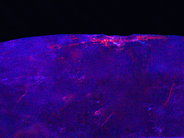
Fourier-transform infrared (FTIR) spectroscopy revealed peaks at 2954, 2924, 2872, and 2854 cm–1 (figure 3), typical of a universal paraffin oil filler (M.L. Johnson et al., “On the identification of various emerald filling substances,” Summer 1999 G&G, pp. 82–107; L. Kiefert et al., “Identification of filler substances in emeralds by infrared and Raman spectroscopy,” Journal of Gemmology, Vol. 26, No. 8, 1999, pp. 501–520). Under deep UV illumination (<225 nm), the foreign filler in the surface-reaching fractures fluoresced strongly pink while the spodumene host rock was inert. The effect was especially prominent in a cavity near the girdle (figure 4).
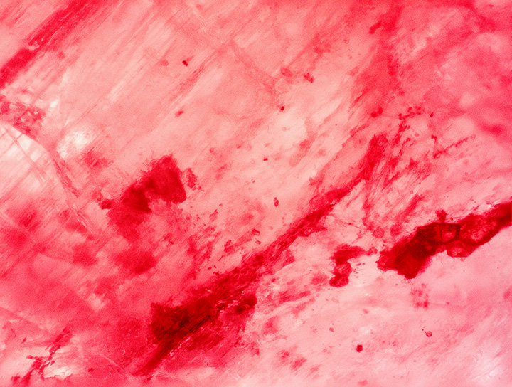
Microscopic examination verified the FTIR, UV-Vis-NIR, and deep UV findings and revealed surface-reaching cleavages and fractures filled with a red foreign substance (figure 5), as well as natural inclusions.
The recorded results confirmed the gem as a red-dyed spodumene. Although spectroscopy identified some manganese, the lack of pink/red reaction under deep UV suggests a low concentration (Lu, 2012). Therefore, the authors assume that the starting material was either colorless or very pale pink spodumene.
Introducing red dye to intensify the color of corundum is a longstanding practice (Schmetzer et al., 1992). To the best of our knowledge, however, this is the first time a spodumene has been treated with a colored oil to imitate ruby. The result masks the true identity of the gem, and without gemological testing, an uneducated trader could misidentify it as a color-enhanced ruby.


