Chart Gems & Gemology, Winter 2018, Vol. 54, No. 4

Chart: Inclusions in Natural, Synthetic, and Treated Diamond
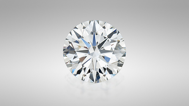

1.15 ct round brilliant diamond
Figure 1. Diamonds such as this 1.15 ct round brilliant are some of the most highly sought-after gem materials and may contain inclusions that give the gemologist clues about their origin or treatment status. Photo by Robert Weldon/GIA.

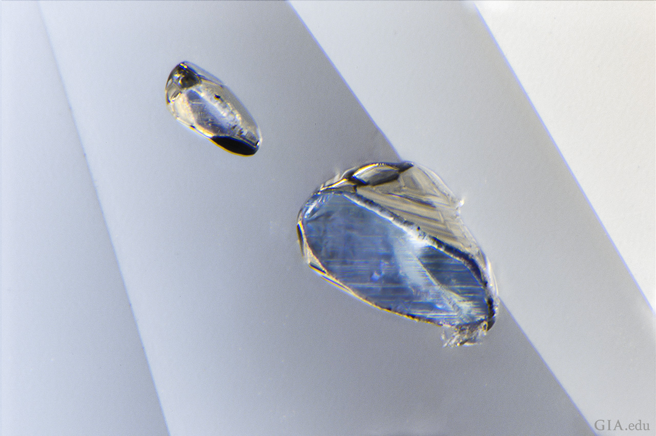
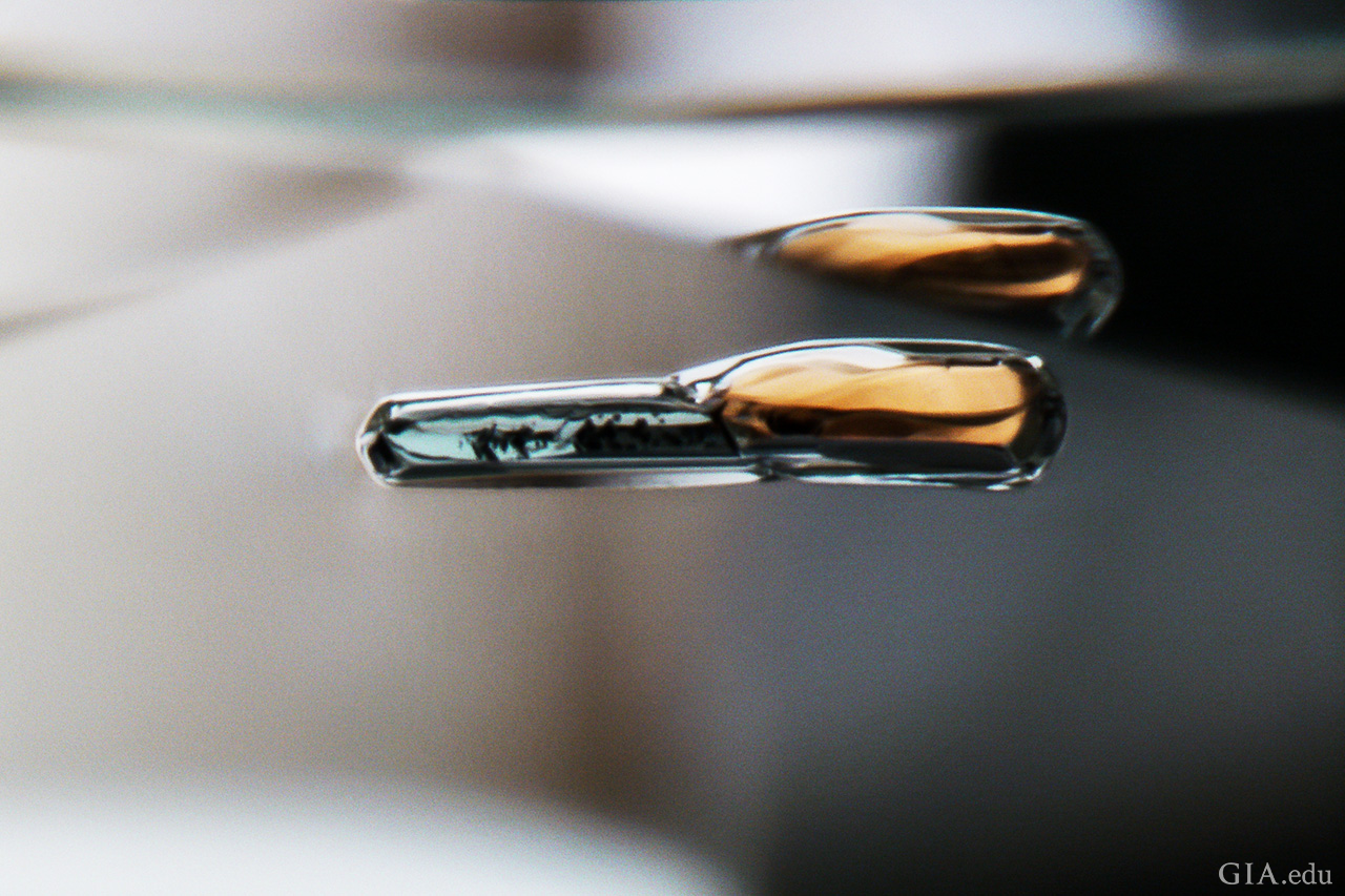
Diamonds (figure 1) are the subject of the latest chart in our series on the micro-features of gemstones. One of the most highly-sought gem materials, diamond—like emerald, sapphire, and ruby—may be treated or grown in laboratory conditions. While many diamonds contain inclusions that are indicative of their origin or treatment status, many gem-quality diamonds are free of such diagnostic characteristics and will require advanced gemological testing to determine whether they are mined, laboratory grown, or treated. For example, chromian garnet inclusions can indicate the diamond formed in a peridotitic environment, while omphacite and kyanite would suggest an eclogitic environment. Other features that are notably diagnostic to the gemologist are so-called “flash-effect” colors that decorate lead glass–filled fractures and tapering tubes that indicate laser drilling.

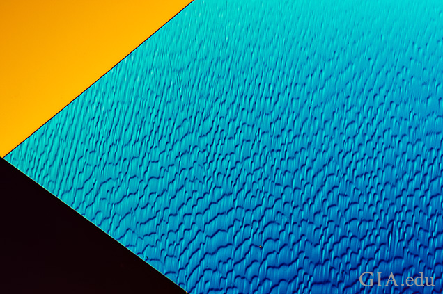
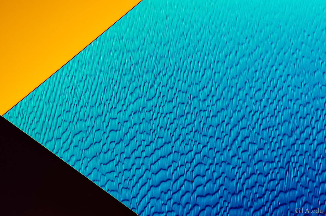
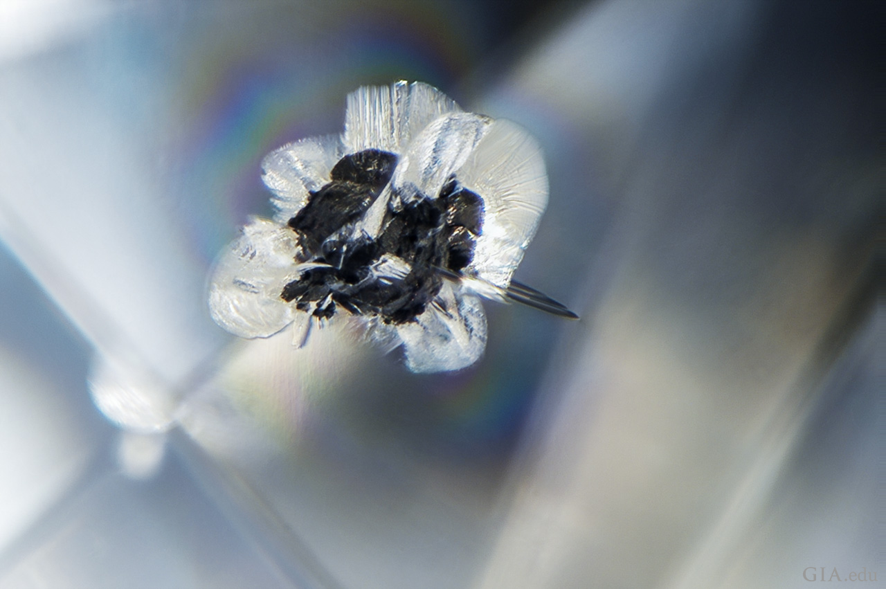
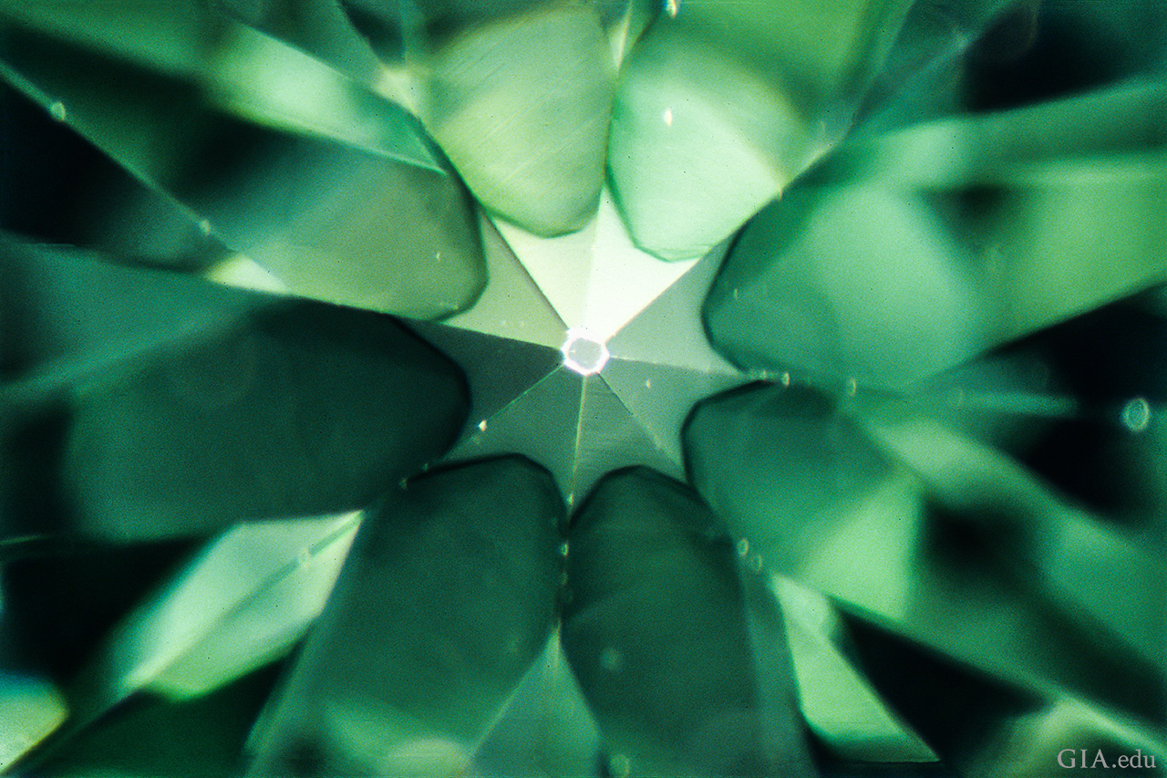
“Lizard skin” results from polishing a facet that lies nearly parallel to the octahedral face, which is the hardest crystal direction in diamond. Field of view 0.72 mm.
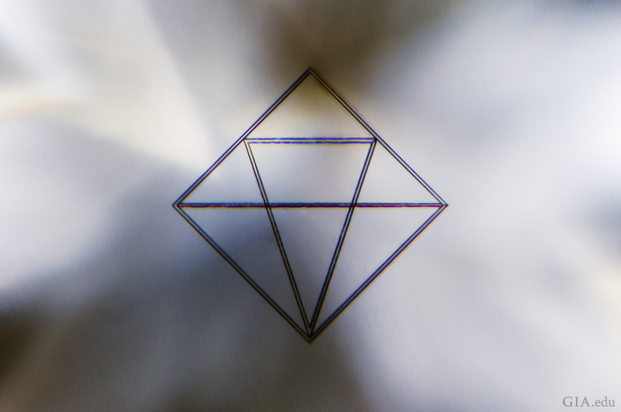
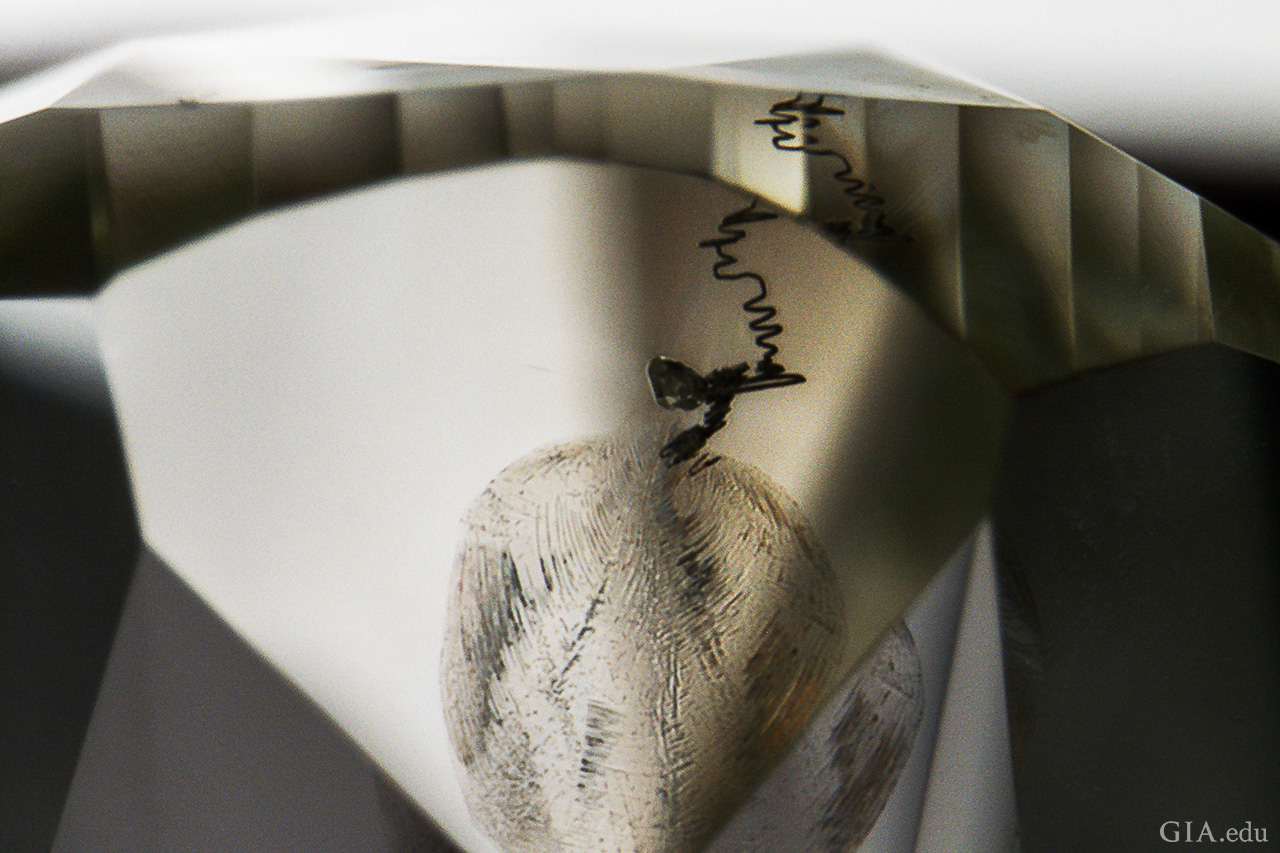
This selection of photomicrographs represent some of the typical and less common inclusions and other growth features seen in natural diamonds.

Gem-quality synthetic diamonds have been commercially available since 1986; however, recent improvements in growth methods are responsible for increases in both the size of finished stones and the quantity produced. Chemical vapor deposition (CVD) and high-pressure, high-temperature (HPHT) processes have had increasing impact in the diamond trade, with CVD stones reaching finished sizes of more than five carats (Eaton-Magaña and Shigley, 2016) and a 15 ct HPHT-grown specimen recently reported (Ardon and Eaton-Magaña, 2018). Melee-size material is also becoming widely available. As a result, GIA offers a melee screening service and has developed a consumer diamond screening device, the iD100, to separate natural and synthetic specimens.

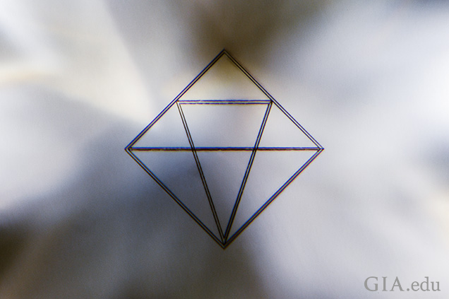
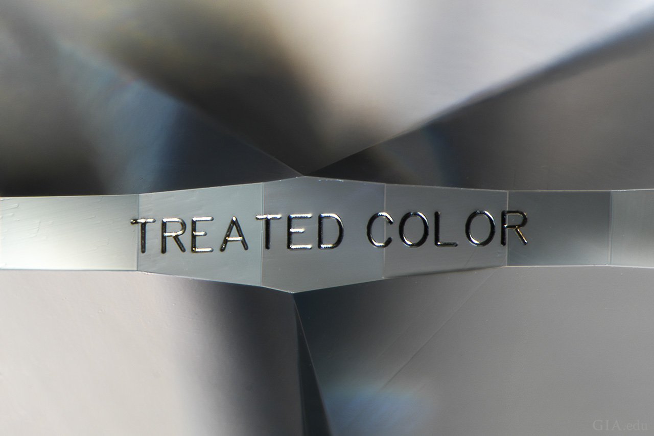
De Beers’ line of CVD laboratory-grown diamonds can be easily recognized by the 300 × 300 micron logo inscribed about 200 microns below the surface of the table facet.
Synthetic diamonds are grown rapidly by two different methods, and they display micro-features that differ from those seen in natural diamonds, which form over much longer periods of time deep in the earth.

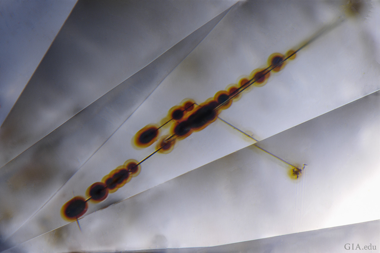
Treatments intended to improve a diamond’s clarity have been around since the late 1970s and include laser drilling and the filling of cracks with a lead glass; this glass reduces the impact of a crack’s appearance on the stone (Koivula et al., 1989). Other treatments may improve or modify the bodycolor of a diamond. Irradiation and HPHT treatment can accomplish this but often leave no microscopic evidence of the process.

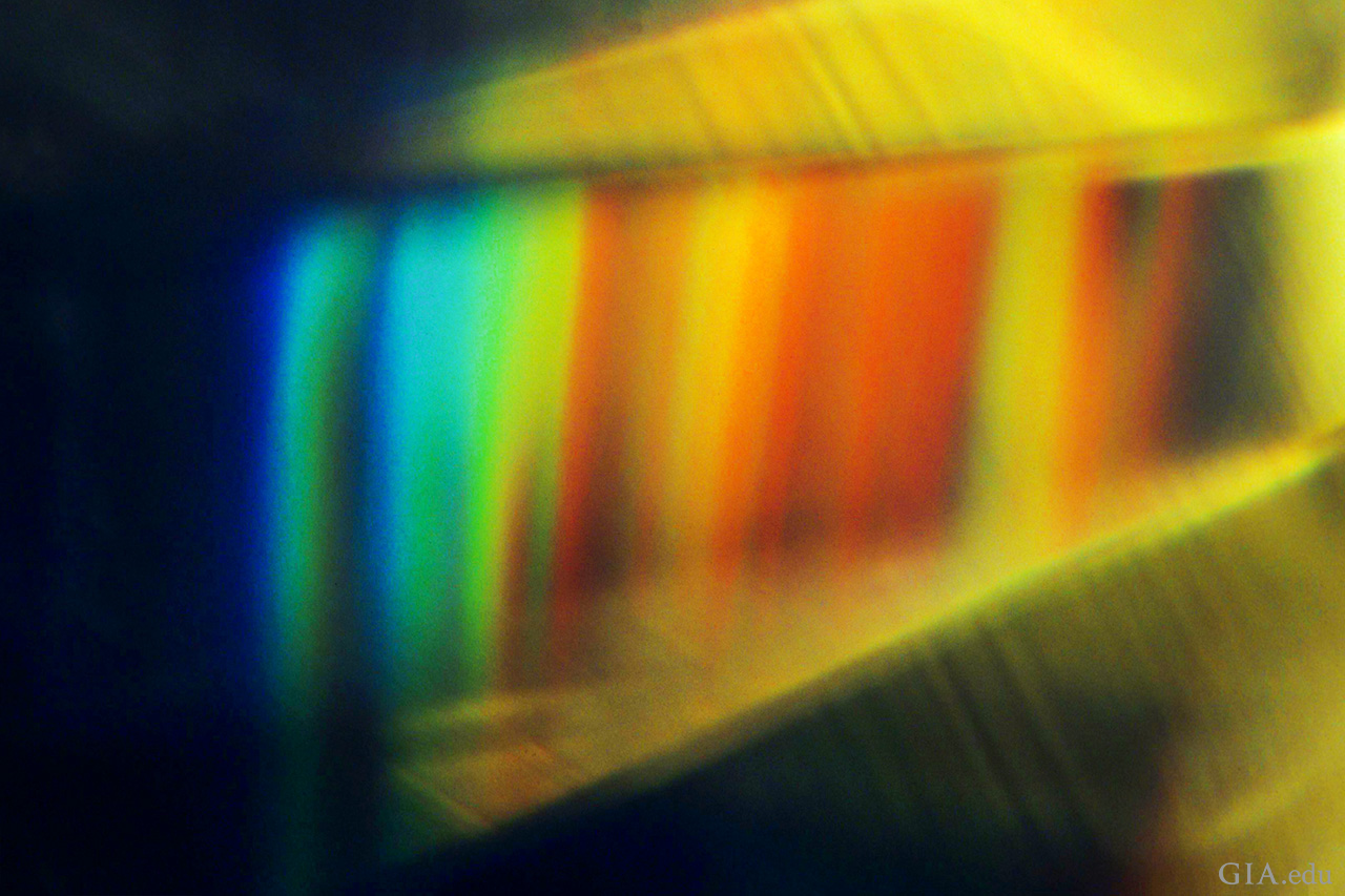
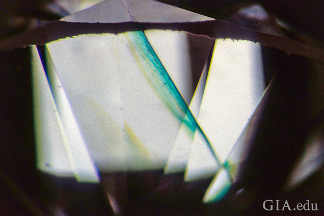
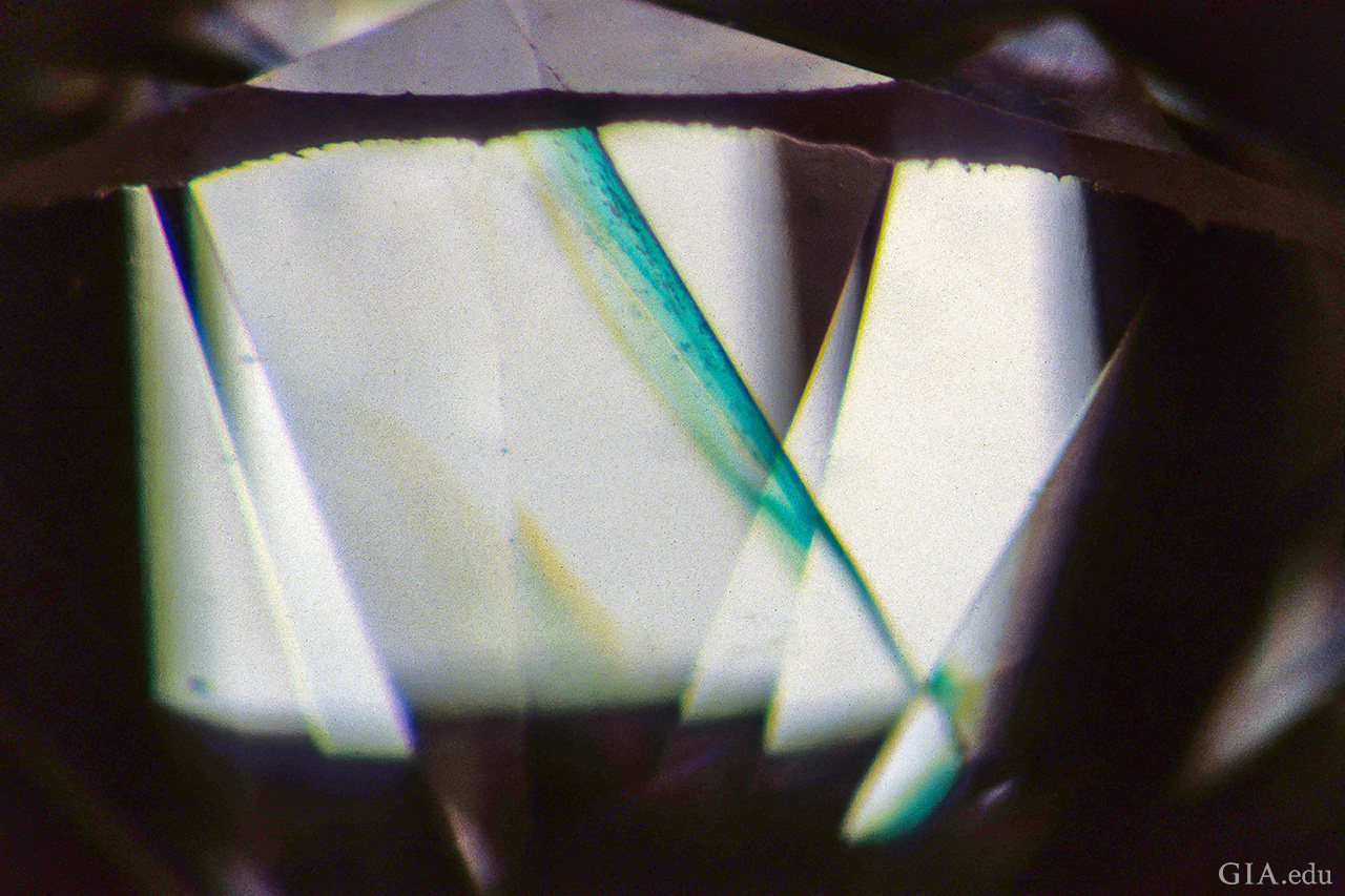
This cleavage crack shows a bright green “flash effect” when observed subparallel to the plane of the break against a bright background. This is diagnostic of clarity enhancement in diamonds. Field of view 3.20 mm.
Diamonds are subjected to treatments to improve their color and/or clarity. The photomicrographs illustrate micro-features often seen in treated diamonds.

While the accompanying chart is not meant to be comprehensive, it aims to remind the gemologist of the microscopic evidence that one may encounter and what that evidence indicates regarding a diamond’s natural, treated, or synthetic origin. For more on diamond and its inclusions, see our suggested reading list.
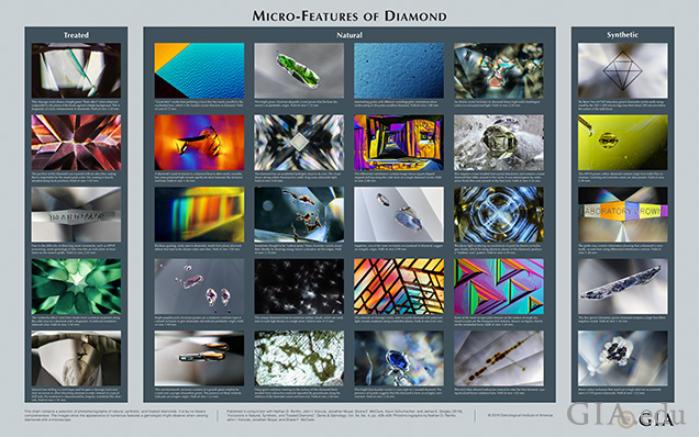
This chart contains a selection of photomicrographs of natural, synthetic, and treated diamonds. It is by no means comprehensive. The images show the appearance of numerous features a gemologist might observe when viewing diamonds with a microscope.
This chart contains a selection of photomicrographs of natural, synthetic, and treated diamonds. It is by no means comprehensive. The images show the appearance of numerous features a gemologist might observe when viewing diamonds with a microscope. Photomicrographs by Nathan D. Renfro, John I. Koivula, Jonathan Muyal, and Shane F. McClure.

Mr. Renfro is manager of colored stones identification, and John Koivula is analytical microscopist at GIA in Carlsbad, California. Mr. Muyal is a staff gemologist, Mr. McClure is global director of colored stone services, Mr. Schumacher is a photo and video producer for Gems & Gemology, and Dr. Shigley is distinguished research fellow at GIA in Carlsbad.

Ardon T., Eaton-Magaña S. (2018) Lab Notes: 15 carat HPHT synthetic diamond. G&G, Vol. 54, No. 2, pp. 217–218.

Eaton-Magaña S., Shigley J.E. (2016) Observations on CVD-grown synthetic diamonds: A review. G&G, Vol. 52, No. 3, pp. 222–245, http://dx.doi.org/10.5741/GEMS.52.3.222

Koivula J.I., Kammerling R.C., Fritsch E., Fryer C.W., Hargett D., Kane R.E. (1989) The characteristics and identification of filled diamonds. G&G, Vol. 25, No. 2, pp. 68–83, http://dx.doi.org/10.5741/GEMS.25.2.68