The Color Origin of Gem Diaspore: Correlation to Corundum

Abstract
Color-change diaspore, known commercially as Zultanite, is sought by designers and consumers for its special optical characteristics, namely its color and color change. Understanding the color origin of gem-grade diaspore could provide a scientific basis to guide its gemological testing, cutting, and valuation. This study uses ultraviolet-visible (UV-Vis) spectra and laser ablation–inductively coupled plasma–mass spectrometry (LA-ICP-MS) to examine the color origin of color-change diaspore and to compare it with corundum. As Raman spectra vibration intensities are closely related to crystal direction for diaspore, crystal orientation was determined through Raman spectroscopy. The color correlation between color-change diaspore and corundum confirmed the identity of each chromophore. In addition, the effectiveness of different chromophores such as Cr3+, Fe3+, Fe2+-Ti4+ pairs, and V3+ between gem-quality diaspore and corundum is compared quantitatively.
Introduction
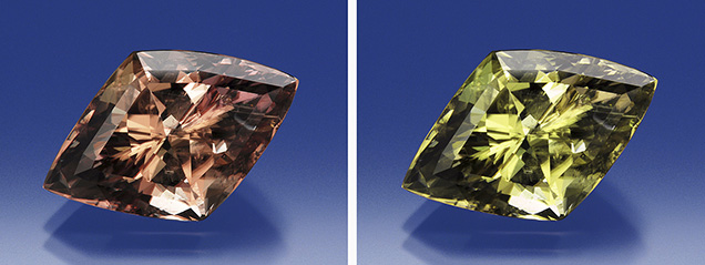
Gem-quality diaspore occupies an important position in the gem market due to its rarity, striking pleochroism, and color-change phenomenon (figure 1). The material’s value depends on these factors. A clear understanding of color origin offers considerable benefits for gemological testing, cutting, and even valuation of gem diaspore.
By replacing the major elements in definite structural units through isomorphous substitution, trace elements play an important role in the color of gemstones. The AlO6 octahedra is a significant structural unit that produces color when different trace elements substitute for Al. For example, Cr3+ substitutes for Al3+ in the AlO6 octahedra in jadeite and spinel, causing green and red color (Lu, 2012; Malsy, 2012), while the substitution of Fe3+ for Al3+ in sapphire produces yellow color (Emmett et al., 2003).

Diaspore and corundum have a similar chemical composition and crystal structure (see figure 2). Diaspore, with the chemical formula AlO(OH), belongs to the orthorhombic space group 2/m 2/m 2/m (Hill, 1979); corundum, with the chemical formula Al2O3, belongs to the trigonal space group 32/m (Lewis et al., 1982). The crystal structure of diaspore consists of AlO4(OH)2 octahedra, whereas the corundum crystal structure consists of AlO6 octahedra (Hill, 1979; Lewis et al., 1982). Both types of crystals are composed solely of octahedral units. In addition, the diaspore structure is able to convert to corundum structure through dehydration (Iwai et al., 1973). Due to their closely related crystallographic structure and chemical composition, we may speculate that there is also a close color correlation. There are two other reasons for this hypothesis:
- Compared to other gemstones containing the AlO6 octahedral structural unit, diaspore and corundum share the closest similarity in average Al-O bond length, octahedral volume, and degree of distortion of octahedral sites (quantified by determining the octahedral quadratic elongation). For specific data, refer to table 1.
- The structural similarity between corundum and diaspore was first referred to by Deflandre (1932). Both diaspore and corundum are connected by octahedral units, and both contain edge-shared octahedra. However, there are face-shared octahedra along the c-axis that only exist in corundum, and this is the main difference between the two. Other related studies involving X-ray powder photography (Francombe and Rooksby, 1959), single-crystal X-ray techniques, scanning electron microscopy (SEM), and high-temperature optical microscopy (Iwai et al., 1973) have confirmed a structural similarity between the two gem materials.

This intensely close relationship between corundum and diaspore with respect to the octahedral Al site and their overall structural similarity indicates that chromophores substituted into the Al site of both materials are expected to present similar UV-visible absorption features. Consequently, this study adopts an analogy to address the color origin of diaspore by quantitatively analyzing the trace-element chemistry in diaspore from two geographic origins—Myanmar (formerly Burma) and Turkey—and comparing them with high-quality natural and synthetic corundum. The research aims to use the color correlation between diaspore and corundum to confirm the color origin of gem diaspore.
MATERIALS AND METHODS
Samples. Nine diaspore samples from Turkey were purchased from a gem dealer at the Beijing International Jewelry Show in November 2016. Forty slices of diaspore were collected in the field from Mogok, Myanmar, by author RL. All samples were prepared as wafers. These wafers are perpendicular to the a-axis, b-axis, and c-axis, respectively. Photos of samples are shown in table 2.

Raman and Photoluminescence (PL) Spectroscopy. Raman and PL spectra were collected from the samples with a Renishaw inVia Raman microscope system at the Gemstone Center of Hebei GEO University in China. Raman spectra were collected from 100 to 1400 cm–1 using Nd-YAG laser excitation, producing highly polarized light at 532 nm (1800 lines/mm grating, 10 s). The orientation of all samples was carefully controlled. The polarization direction of the injected laser was parallel to the a-axis, b-axis, and c-axis. The 600–800 nm spectral ranges for Raman photoluminescence analysis (under liquid nitrogen conditions) were excited by an Nd-YAG laser at 532 nm (1800 lines/mm grating, 10 s).
LA-ICP-MS Analysis. For trace-element analysis, we used an Agilent 7900a ICP-MS coupled with a deep-UV laser at 193 nm excitation at the State Key Laboratory of Geological Processes and Mineral Resources, China University of Geosciences in Wuhan. NIST glass standard SRM 610 and USGS glass standards BHVO-2G, BIR-1G, and BCR-2G were used for external calibration. Ablation was achieved using a 44 μm diameter laser spot size, a fluence of around 5 J/cm2, and a 6 Hz repetition rate. The diaspore was initially standardized internally with 27Al using a fixed internal standard method. We selected three spots on each sample for general chemical composition.
UV-Vis Spectroscopy. Five diaspore samples (Dia-006-a, Dia-006-b, Dia-006-c, Dia-Bur-001, and Dia-Bur-002) were prepared as well-polished, oriented wafers with various thicknesses. UV-Vis spectra were collected with a Perkin-Elmer Lambda 650 UV-Vis spectrophotometer equipped with mercury and tungsten light sources and photomultiplier tube (PMT) detectors installed in an integrating sphere. The spectra were collected at the Gemological Institute, China University of Geosciences in Wuhan. Polarized spectra were collected in the 300–800 nm range with 1 nm spectral resolution at a scan speed of 267 nm/min. The spectral baseline was corrected by subtracting spectral offsets at or beyond 800 nm, where the chromophores’ features were insignificant or nonexistent.
RESULTS AND DISCUSSION
Chemical Analysis. Table 2 shows the chemical composition (expressed in ppma) of two diaspore samples from Turkey (Dia-006, Dia-008) and two from Myanmar (Dia-Bur-001, Dia-Bur-002). We concluded that the Burmese samples contained more Cr, while the Turkish diaspore had a higher Fe content. However, the V content of the Burmese samples was higher than that of the Turkish samples, especially in Dia-Bur-002 (358 ppma).
Raman Spectroscopy. Unlike Turkish diaspore, Burmese material often occurs as thin crystals that are difficult to orient by crystal growth characteristics. However, Raman vibration is closely related to crystal direction (see http://rruff.info/diaspore/display=default/R060287). By comparing the Raman spectra of the diaspore from Myanmar with the oriented sample from Turkey, the Burmese diaspore could be oriented properly. Raman spectra of both the Burmese and Turkish diaspore were collected in the 100–1400 cm–1 range.
According to previous research by Ruan et al. (2001) and San Juan-Farfán et al. (2011), the band at 154 cm–1 is assigned by the rotation of two edge-shared AlO6 octahedra around the c-axis. The most intense peak, at 447 cm–1, is related to Al-O symmetric stretching modes, which means the different polarization direction of the laser has little impact on this peak. Therefore, we normalized the peak intensity at 447 cm–1 and compared the intensity at 154 cm–1. When the polarization direction of the injected laser is parallel to the c-axis, the intensity of the 154 cm–1 peak is much stronger than those that run parallel to the b-axis and a-axis (see note on figure 3). Based on figure 4, we can accurately diagnose the crystal orientation by Raman vibration (154 cm–1). The Burmese diaspore wafers were manufactured perpendicular to the b-axis based on perfect cleavage along the {010} direction. Hence, we can consider the long side of the wafer parallel to the c-axis, and the short side parallel to the a-axis.


UV-Vis and PL Spectroscopy of Cr3+. Corundum, a uniaxial crystal, shows dichroism. Its UV-Vis spectra are often collected from two orthogonal orientations with polarized light (o-ray and e-ray). As a biaxial crystal, diaspore shows trichroism; its UV-Vis spectra are collected from three orthogonal orientations with polarized light (E ǁ a, E ǁ b, and E ǁ c). In gem cutting, the table of a ruby or sapphire is generally perpendicular to the c-axis. When the polarization direction (E vector) is parallel to the a-axis, diaspore shows UV-visible absorption features similar to those of corundum (see figure 5). Our corundum samples included three synthetic sapphires—a Cr-bearing synthetic ruby, a Fe-Ti bearing synthetic blue sapphire, a V-bearing color-change sapphire—and one natural yellow Fe-bearing sapphire from Garba Tula, Kenya.

In order to make our initial comparisons between chromophores in corundum and diaspore, we have chosen to compare the spectra of corundum collected from the o-ray (R. Lu, previously unpublished data) with those of diaspore collected from the orientation of polarized light parallel to the a-axis. From LA-ICP-MS data, we know that the Cr content is significantly higher in Burmese samples than in Turkish samples. However, as the Fe content is near zero in the Burmese samples, we disregarded the absorption of Fe in the UV-Vis spectra of that material. The spectra of Dia-Bur-001 and Dia-Bur-002 are shown in figure 6. Although the Cr content of the two samples is almost the same (2166 and 2081 ppma, respectively), there is a large difference in the absorption intensity. This difference is related to the absorption of vanadium. By subtracting the two spectra, we essentially obtain only V absorption, which can be subtracted from the other spectra to obtain the pure Cr spectrum (see box A). Based on the author’s color analysis, chromium will cause a slight color-change phenomenon in diaspore (figure 7). Additionally, we used the PL spectra (figure 8) to confirm that gem diaspore and ruby show comparable fluorescence spectra (694/693 nm for ruby and 690/693 nm for diaspore).


UV-Visible Spectroscopy of Fe3+ in Diaspore and Sapphire. The UV-Vis spectrum of Turkish diaspore (figure 9) shows features corresponding with those of Fe3+ -bearing yellowish sapphire from Garba Tula, Kenya. In those sapphires, the peak at 388 nm is attributed to Fe3+, while the peaks at 377 and 450 nm are attributed to Fe3+-Fe3+ pairs (Ferguson and Fielding, 1971, 1972; Krebs and Maisch, 1971). The spectra of diaspore show features very similar to those of Fe3+ -bearing sapphire. Hence, the peaks at 384 and 448 nm are attributed to Fe3+-Fe3+ pairs, and the peak at 398 nm is attributed to Fe3+.

UV-Visible Spectroscopy of Fe2+-Ti4+ Pairing in Diaspore and Sapphire. The UV-Vis spectra of the Turkish diaspore show an obvious absorption band at around 570 nm. Based on the LA-ICP-MS data, chromium and vanadium are very low in Turkish diaspore. In blue sapphire, the absorption at around 580 nm is attributed to an Fe2+-Ti4+ pairing (Emmett et al., 2003). It is possible that the absorption at around 570 nm in diaspore is related to Fe2+-Ti4+ (see figure 10). In blue sapphire, the absorption of the Fe2+-Ti4+ pair does not cause the color-change effect because the absorption band extends to the near-infrared region, so the transmission rate of red areas and green areas is not relatively equal. But in diaspore, these transmission areas are fairly equal. The value of a* has changed from –3 to 1, which means the color will change from green to red.

Emmett et al. (2017) proposed that Si plays a role in the color chemistry of corundum. If corundum contains Ti, Si, Mg, and Fe, the Fe will pair with Si before Ti. If diaspore is similar to corundum, Fe will charge-compensate Si before Ti. However, the instruments used for LA-ICP-MS show significant interferences for the three silicon isotopes (Shen, 2010), and as a result the Si content is higher than the value shown. The author does not take Si into consideration because of this instrumental error, and assumes that all of the Ti pairs with Fe. Therefore, the actual content of the Fe2+-Ti4+ pair may be lower than the content determined through analysis.
UV-Visible Spectroscopy of V in Diaspore and Sapphire. In natural corundum, vanadium content is generally very low. Synthetic sapphire, which has been manufactured with V, will show an obvious color-change effect. This type of synthetic sapphire is similar to natural alexandrite. However, there is some V in natural diaspore from Myanmar, which plays an important role in color origin and color change.
We compared the spectra of diaspore (containing V only; see box A) to pure vanadium-bearing synthetic sapphire. They also show similar absorption characteristics in their UV-Vis spectra, as both peaks are at around 400 nm and 560 nm in diaspore and sapphire (see also figure 11).

CONCLUSIONS
In color-change diaspore, Cr3+, V3+, and Fe2+-Ti4+ pairs are the chromophores, and all of these contain an absorption area at around 560–580 nm. These chromophores may all play a role in causing the color-change effect. Raman spectroscopy proved to be a powerful tool for determining the crystal orientation of the Burmese diaspore wafer; it is also useful to measure and compare the directional UV-Vis spectra of diaspore and corundum. Because of their structural similarity, we can confirm the color correlation between corundum and diaspore and compare the effectiveness of the chromophores. According to the calculation, the chromophore effectiveness of Cr3+, Fe3+, and Fe2+-Ti4+ in corundum is about 5–10, 1.6, and 50 times higher, respectively, than in diaspore. However, the chromophore effectiveness of V3+ in diaspore is approximately 2–7 times higher than in corundum.


