Uranium Contents of Hyalite
Hyalite is a colorless variety of common opal with strong greenish fluorescence. Rough specimens have interesting glassy botryoidal shapes formed by vapor transport in volcanic or pegmatitic environments (O.W. Flörke, “Transport and deposition of SiO2 with H2O under supercritical conditions,” Kristall und Technik, Vol. 7, 1972, pp. 159–166). Hyalite from a new source in Zacatecas, Mexico, discovered in 2013, shows strong green fluorescence even under daylight conditions. This material was spotlighted for the first time at the 2014 Tucson gem and mineral shows (E. Fritsch et al., “Green-luminescing hyalite opal from Zacatecas, Mexico,” Journal of Gemmology, Vol. 34, 2015, pp. 490–508).
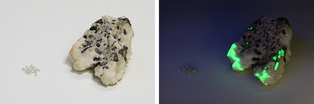
GIA’s Tokyo laboratory recently had an opportunity to examine several faceted hyalites from Zacatecas, Mexico, along with several rough hyalites from other locations, including Japan, Hungary, and Argentina. There were two different types of Japanese hyalites in the study samples: botryoidal and spherical. The botryoidal one (figure 1) was from Gifu’s Naegi granitic pegmatite (H. Ogawa et al., “Fluorescence of hyalite in pegmatite from Naegi granite, Nakatugawa,” 2008 Annual Meeting of Japan Association of Mineralogical Sciences), while the spherical one was from Toyama’s Shin-yu hot spring (Y. Takahashi et al., “On the occurrence of opal at the Shin-yu hot spring, Tateyama,” Tateyama Caldera Research, Vol. 8, 2007, pp. 1–4.). Hungarian hyalite is known to form in the clefts of trachytic rocks in Bohemia, Czech Republic, and Auvergne, France (B. von Cotta, Rocks Classified and Described: A Treatise on Lithology, Longmans, Green, and Company, London, 1866, p. 425). Argentinean hyalite is widely distributed, but the original rocks are not described.
Standard gemological testing of the faceted Mexican stones revealed RI values of 1.460–1.461 and a hydrostatic SG of 2.13. Prominent fluorescence was observed under short-wave UV light, but only the Japanese spherical samples were inert (figure 2). Strong graining flow structures were visible at 64× magnification. Raman analysis (figure 3) revealed typical amorphous silica spectra composed of a major band between 430 and 460 cm–1 (vibration of bridging oxygen) and a band centered at 790 cm–1 representing the stretching of isolated SiO44– (P. McMillan, “A Raman spectroscopic study of glasses in the system CaO-MgO-SiO2,” American Mineralogist, Vol. 69, 1984, pp. 645–659). Laser ablation–inductively coupled plasma–mass spectrometry (LA-ICP-MS) detected a very wide range of uranium contents; the maximum was 1060–1330 ppmw for the Japanese pegmatitic hyalite, while the Japanese spherical hyalite had zero uranium content. The amounts are quite inhomogeneous, but seem to correlate with the intensities of fluorescence, decreasing from Mexican (1180–3.55 ppmw) to Argentinian (2.48–2.49 ppmw) to Hungarian (0.39–0.15 ppmw) (figure 3).


Interestingly, only the Japanese pegmatitic hyalite sample was rich in rare earth elements (REE). Since Naegi granitic pegmatite is known to be an REE-enriched pegmatite (T.S. Ercit, “REE-enriched granitic pegmatites,” in R.L. Linnen and I.M. Samson, Eds., Rare-Element Geochemistry and Mineral Deposits, Geological Association of Canada, GAC Short Course Notes 17, 2005, pp. 175–199), the host rock chemistry appears to affect the silica-rich fluid.


