Natural Pearl Aggregates from Pteria Mollusks
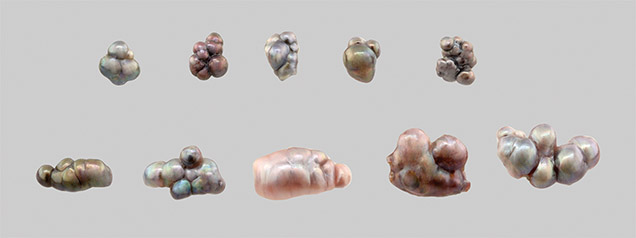

Figure 2. Microradiograph images of the pearls revealed multiple growth units with central conchiolin-rich cores in each unit. Images by Yixin (Jessie) Zhou.
Microscopic examination revealed that the pearls formed with continuous nacre layers. Microradiography further demonstrated that their internal structures were composed of multiple small pearls related to their growth (figure 2). Conchiolin-rich centers were observed in all of the sub-units, indicating natural formation. Additional advanced testing (UV-Vis reflectance spectrophotometry, photoluminescence spectroscopy, and EDXRF spectrometry) showed that all 10 of the pearls had a natural color. More importantly, all 10 exhibited medium to strong orangy red to red fluorescence under long-wave UV, which is characteristic of pearls that form in Pteria mollusks (figure 3).
Figure 3. The pearls produced this striking characteristic red fluorescence under long-wave UV. Photo by Jian Xin (Jae) Liao.
Pearls grown from the aggregation of smaller pearls are sometimes associated with freshwater culturing practices (Summer 2012 Lab Notes, pp. 138–139). Yet these samples clearly did not appear to be cultured. Our client later informed us that the pearls were obtained more than 40 years ago, reportedly off the Mexican coast, which further supports the identity of these unique pieces.


