Calcite Pearl with Red X-Ray Fluorescence

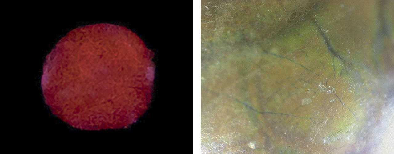
GIA’s Mumbai laboratory recently received for identification seven strands containing 1,170 variously shaped white to cream-colored pearls. Real-time X-ray microradiography (RTX) revealed that most of the pearls were of natural origin, and a few were non-bead cultured pearls. All the natural pearls showed an inert reaction when exposed to X-ray excitation due to their saltwater origin, except for one white near-round pearl that measured 2.55 mm in diameter and weighed approximately 0.12 ct (figure 1). The pearl exhibited a very unusual deep red fluorescence (figure 2, left).
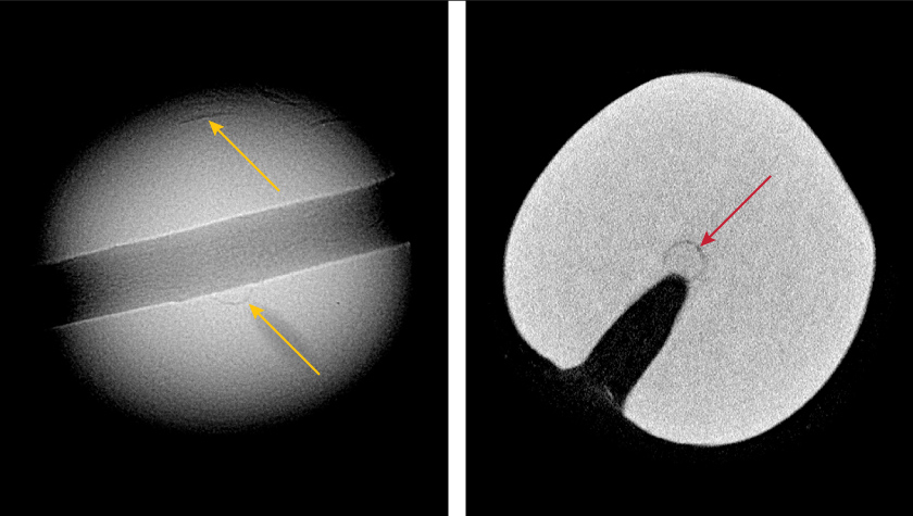
RTX analysis showed a minimal growth structure with a few faint growth arcs in the outer area of the pearl and one distinct growth ring closer to the center near the drill hole (figure 3, left). X-ray computed microtomography (μ-CT) further revealed a very fine growth arc within the center ring (figure 3, right). These were consistent with structures observed in natural pearls from the Pinctada species.
Externally, the pearl looked similar to the other pearls in the strands, possessing a medium surface luster with orient. Examination at high magnification exposed a translucent layer showing characteristic fingerprint-like overlapping surface platelets with blue subsurface vein-like features branching out in a dendritic pattern beneath it (figure 2, right). Interestingly, similar blue vein-like features have been observed in some research pearl samples reportedly produced from Pinctada radiata mollusks from the Persian (Arabian) Gulf. No visual indications of treatment on the surface were observed. Under long-wave ultraviolet light, the pearl exhibited a moderate chalky bluish green reaction. Energy-dispersive X-ray fluorescence revealed a manganese level of 76 ppm and a strontium level of 1599 ppm, characteristic of saltwater origin.
Raman analysis conducted on two randomly chosen spots using 514 nm laser excitation revealed peaks at 154, 282, 712, 1085, and 1437 cm–1, indicating calcite composition. Saltwater calcite pearls with a “nacreous-looking” surface have been previously examined by GIA laboratories (Winter 2022 Lab Notes, pp. 477–478; Spring 2024 Lab Notes, pp. 69–71). However, they lacked visible fluorescence to X-ray excitation and exhibited a clear, evenly spaced fine concentric ring structure, contrasting with the minimal growth structure of this pearl.
Spectroscopic analysis combined with microscopic examination suggested that the red fluorescence was not a result of any surface treatment, but rather a natural phenomenon. Orange to red fluorescence has been previously recorded in freshwater pearls containing calcite and vaterite (both of which are polymorphs of calcium carbonate) when activated by cathodoluminescence or X-ray excitation. This luminescence has been associated with the presence of Mn2+ in either calcite or vaterite in freshwater non-bead cultured pearls from China (S. Karampelas et al., “Chemical characteristics of freshwater and saltwater natural and cultured pearls from different bivalves,” Minerals, Vol. 9, No. 6, 2019, article no. 357). Although such red fluorescence in saltwater pearls has not been previously recorded, we suspect a connection to the presence of calcite on the pearl’s surface, though the existence of vaterite in the inner nacre layers remains a possibility.
Further research on the chemical composition of the surface and inner cross-sectional layers of this sample is required to understand the exact formation and the specific cause of this reaction in saltwater pearls. This is the first time GIA has encountered a nacreous-looking calcite pearl that exhibited red fluorescence when subjected to X-ray excitation.


