A Fascinating Large South Sea Bead Cultured Pearl

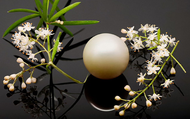
A recent noteworthy submission to GIA’s Mumbai laboratory contained a loose round, undrilled white pearl with strong orient, weighing 54.96 ct and measuring 19.90 × 19.53 mm (figure 1). Externally the pearl had a smooth, blemish-free surface and exhibited a homogeneous uniform color with a high luster. When viewed under 40× magnification, large and evenly spaced overlapping aragonite platelets were visible which helped account for the silky reflection on the surface. X-ray fluorescence (XRF) analysis gave a relatively inert reaction. Chemical analysis using energy-dispersive X-ray fluorescence spectrometry was performed on two surface areas. The data collected showed manganese levels on both areas below the detection level of the instrument and higher strontium levels of 1162 and 1163 ppm, confirming the pearl formed in a saltwater environment. A moderate greenish yellow reaction was observed under long-wave ultraviolet radiation, and a much weaker greenish yellow reaction was noted under short-wave UV.

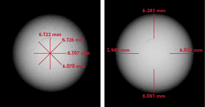
Due to the pearl’s large size, it was quite intriguing to see the growth structure revealed from within. Real-time microradiography (RTX) imaging showed an extremely small bead nucleus together with distinct growth arcs flowing in an even pattern around the nucleus proving it to be a bead cultured (BC) pearl. The exceptional thickness of the nacre growth around the bead was particularly notable (figure 2) (L.M. Otter et al., “A look inside a remarkably large beaded South Sea cultured pearl,” Spring 2014 G&G, pp. 58–62). The size of the bead and the nacre thickness were measured using an option available on the RTX instrument, resulting in a reading of 6.122 mm for the thickest part of the bead and a range of 5.987 to 6.283 mm, in four different directions, for the overlying nacre. These measurements indicated a composition of more than 96% nacre and less than 4% bead nucleus by volume.

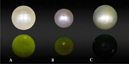
The majority of beads used in the cultivation process of BC pearls are produced from freshwater mollusk shell (Unionidae) collected in the United States (A. Homkrajae et al., “Internal structures of known Pinctada maxima pearls: Cultured pearls from operated marine mollusks,” Fall 2021 G&G, pp. 186–205). Due to the high levels of manganese present in freshwater mollusks, such BC pearls would emit a weak or moderate yellowish green fluorescence when subjected to XRF analysis. This reaction is normally based on the size of bead nuclei used in the cultivation process and the thickness of the surrounding nacre. Hence, the reaction observed via XRF for the pearl was relatively weak due to the thickness of its nacre overgrowth (figure 3) (S. Karampelas et al., “Chemical characteristics of freshwater and saltwater natural and cultured pearls from different bivalves,” Minerals, Vol. 9, No. 6, 2019, article no. 357). Taking all these factors into consideration—external appearance, dimensions, size of the bead nucleus, nacre thickness measurements, and chemistry data—the pearl was classified as a BC pearl produced from the Pinctada maxima species.
Over the decades, South Sea BC pearls have played an important part in the global pearl trade. Larger pearls, some more than 20 mm, were also produced, but these are very rare even with today’s modern farming methods. Their uniquely large size, silky luster, and greater nacre thickness compared with BC pearls from other mollusks make them more attractive and durable. This has allowed them to establish a prominent position in the cultured pearl value chain as the primary choice of consumers looking for large white to cream, or “golden” BC pearls. As a result, they have been used by renowned jewelry designers in their exclusive collections. GIA continues to test numerous South Sea cultured pearls, and this unique submission to the Mumbai laboratory was particularly notable for its size and fine quality.


