Quench-Crackled Dyed Blue Chalcedony Resembling Larimar
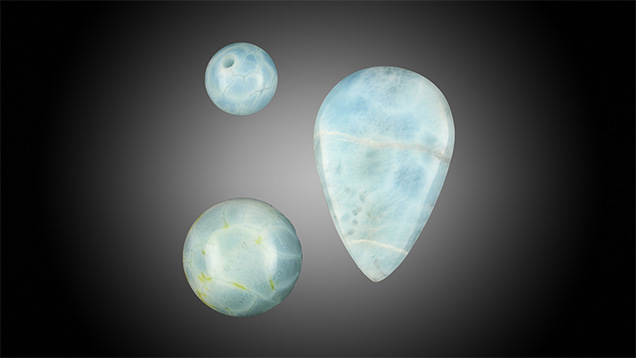
Recently, GIA’s Tokyo laboratory examined five round beads that were semitransparent to opaque and showed blue to greenish blue bodycolors with a network-like structure of whitish zones (figures 1 and 2). These beads were acquired at an ornamental gem material shop in Tokyo and were sold as “sea-blue chalcedony,” but the resemblance to Larimar led to our investigation.

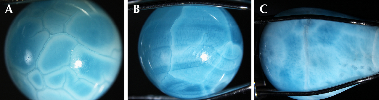
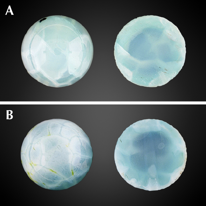
The beads were identified as chalcedony based on spot RI readings of 1.53 to 1.54, SG values ranging from 2.48 to 2.65, and microscopic features such as parallel curved bands (figure 2B). Raman spectroscopic features (figure 3) also suggested that the beads were mainly composed of quartz crystals and matched chalcedony (e.g., D. Pop et al., “Raman spectroscopy on gem-quality microcrystalline and amorphous silica varieties from Romania,” Studia Universitatis Babeș-Bolyai, Geologia, Vol. 49, 2004, pp. 41–52). Raman spectra of the beads showed no characteristics of chrysocolla (Spring 2020 Gem News International, pp. 188–189). Qualitative EDXRF analysis detected peaks related to Si, Fe, and Cu, indicating that Cu was likely the color-causing element. Ultraviolet-visible (UV-Vis) spectra also showed a broad band from 500 to 1000 nm that corresponded with Cu. There was no polymer detected by Fourier-transform infrared (FTIR) spectroscopy and no reaction when touched with a hot point. Under a gemological microscope, the stones revealed whitish zones following fractures. In order to consider the overall structure of such fractures, we cut them in half. Cross sections of the beads showed that the fractures with whitish zones were concentrated on the surface (figure 4). Such a structure is a known characteristic of quench-crackled dyed chalcedony and was reported previously (see Winter 2009 Lab Notes, p. 288). These beads probably represent a new color variety.
These quench-crackled dyed chalcedonies resemble Larimar in color and appearance. Larimar is a rare blue variety of pectolite with the ideal chemical formula of NaCa2Si3O8(OH) that displays white, green, and pale to sky blue colors. Blue color seen in Larimar is believed to be caused by the presence of small amounts of Cu2+ within its structure (e.g., R.E. Woodruff and E. Fritsch, “Blue pectolite from the Dominican Republic,” Winter 1989 G&G, pp. 216–225). We compared these beads with Larimar (the pear cabochon on the right in figure 1) to reveal their differences. The Larimar cabochon showed a mottled blue and white color with network-like whitish zones and other natural mineral inclusions (figure 2C) (again, see Woodruff and Fritsch, 1989). It had a spot RI reading of 1.60 and an SG of around 2.80. On the other hand, the quench-crackled dyed blue chalcedony beads had whitish zones following a network of fractures (figure 2B) and no other mineral inclusions.
This material can easily be separated from Larimar using standard gemological testing such as RI and SG. Advanced testing is useful in confirming the cause of blue color and the presence of polymer treatment.


