Feature Gems & Gemology, Summer 2019, Vol. 55, No. 2

Inclusions in Natural, Treated, Synthetic, and Imitation Opal
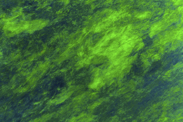

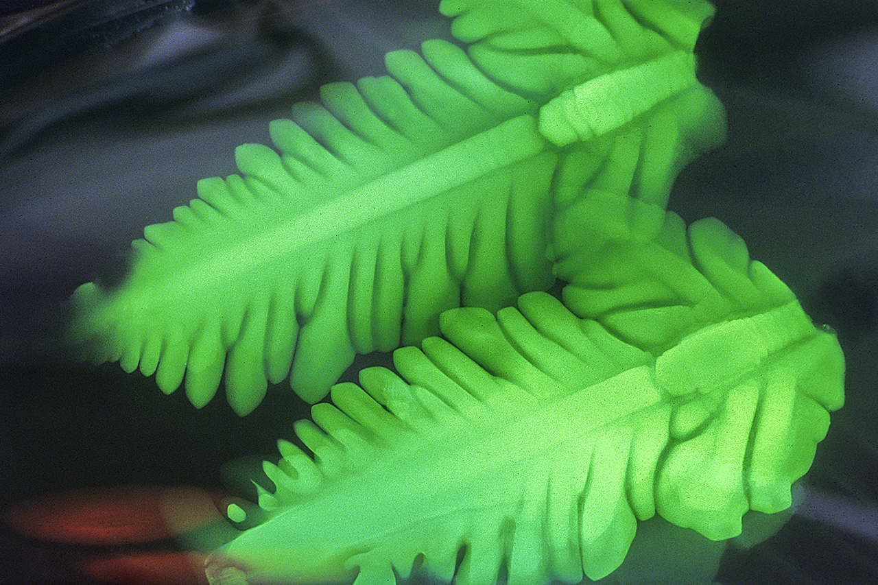
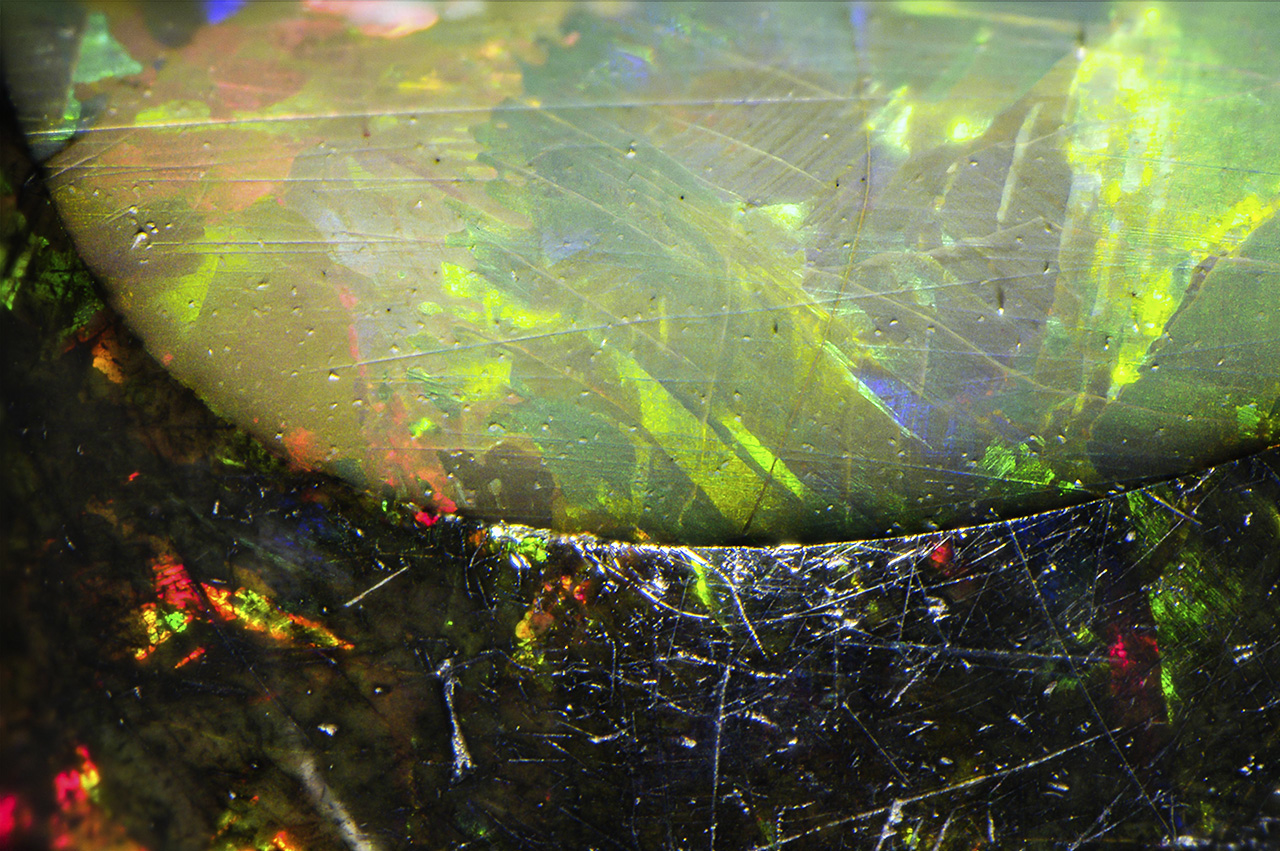
Play-of-color is the defining characteristic of gem opals. While play-of-color can be quite spectacular under magnification, as with the roiled green patterns seen in this crystal opal, other microscopic features are often found if one takes the time to look. Field of view 9.60 mm. Photomicrograph by Nathan Renfro
Figure 1. Play-of-color is the defining characteristic of gem opals. While play-of-color can be quite spectacular under magnification, as with the roiled green patterns seen in this crystal opal, other microscopic features are often found if one takes the time to look. Field of view 9.60 mm. Photomicrograph by Nathan Renfro.

When gemologists think of opals, play-of-color is almost certainly the first characteristic that comes to mind (figure 1). While play-of-color patterns can be extraordinarily beautiful under magnification, opals often contain a vast array of spectacular microscopic features in addition to this phenomenon. In continuing G&G’s series on inclusions, this chart will focus on natural, treated, synthetic, and imitation opals.

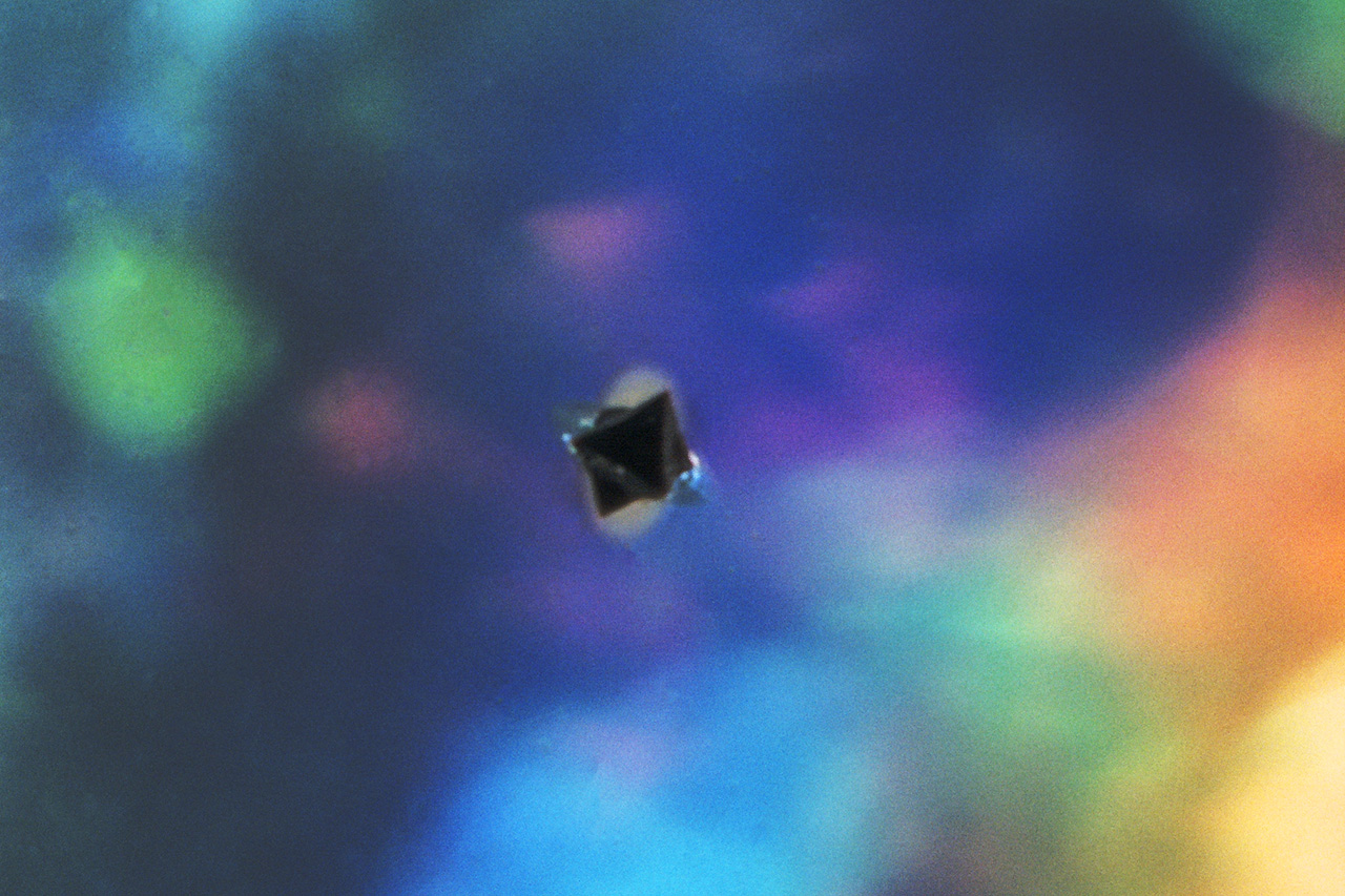
There have been significant developments in the opal industry in recent years with the discovery of opal from Wollo Province in Ethiopia (Rondeau et al., 2010). This deposit produces opals that contain a wide range of interesting inclusions such as fossilized plant material. Opals from Australia also have the potential to showcase spectacular inclusion scenes, from pyrite crystals to black plumes of manganese oxide that stand out in high contrast to the play-of-color phenomenon. Other sources of natural opal that contain visually striking inclusions are Mexico, the United States, Honduras, and Peru.

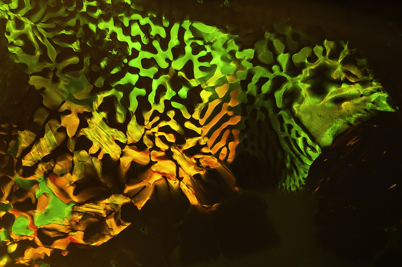
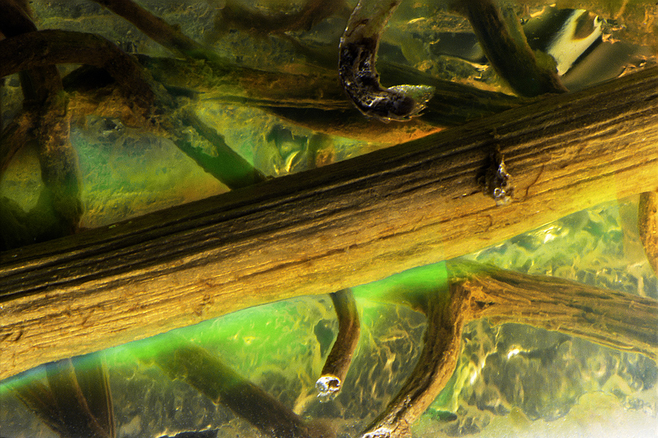
Viewed perpendicular to the grain of this opalized wood from Virgin Valley, Nevada, the cellular structure of the wood shows up as distinct linear strings of play-of-color spots. Field of view 4.87 mm.
Natural opals can occasionally display amazing colors and structure patterns that are the result of their geological history of formation.

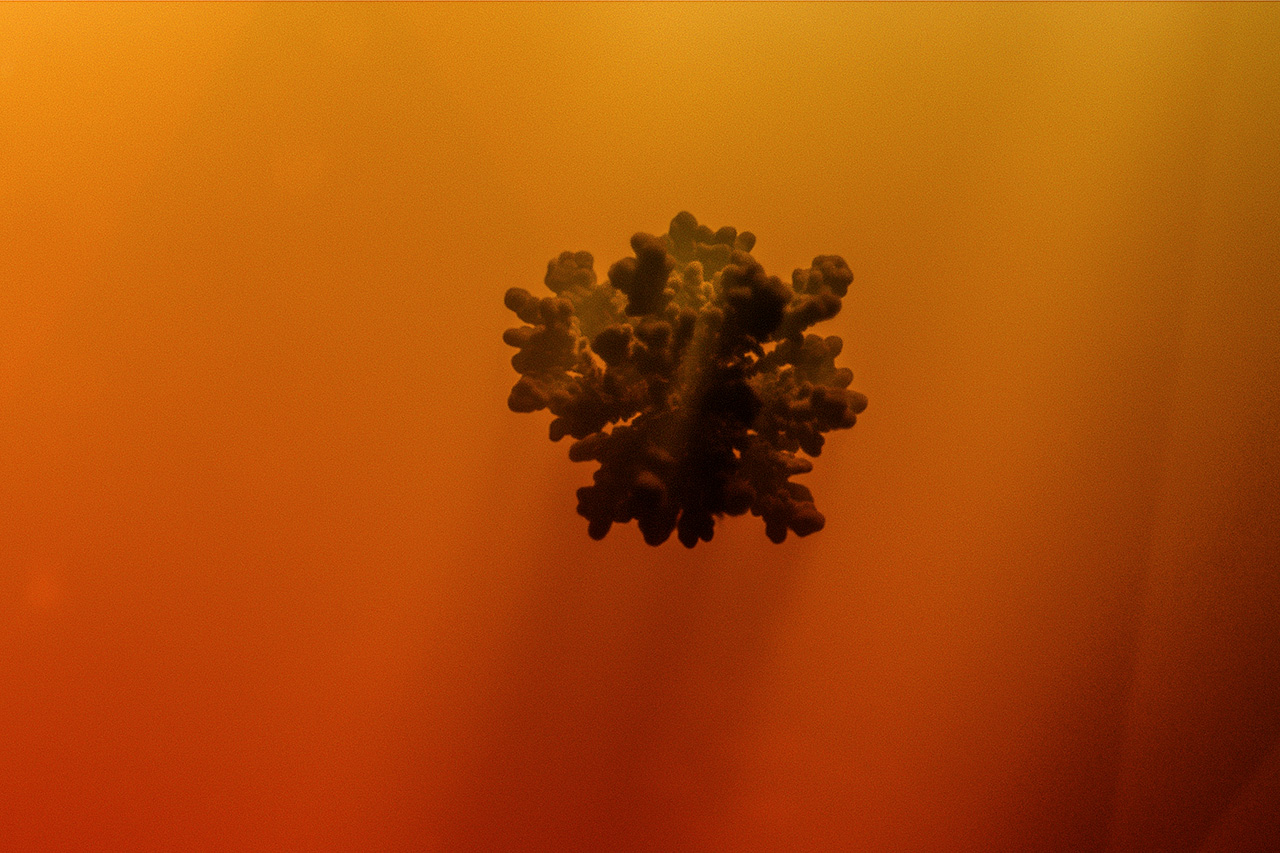
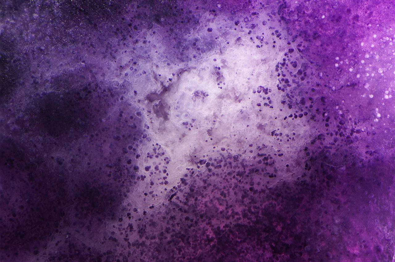
Along with the new Ethiopian deposit came new treatments. The opal from this deposit was hydrophane, which meant it would readily absorb liquids. Soon gem treaters took advantage of this property and began to dye Ethiopian opal a myriad of colors, most notably purple (Renfro and McClure, 2011). This material also proved quite responsive to smoke treatment, which gives it a dark bodycolor (Williams and Williams, 2011). Other traditional opal treatments include color modification by sugar treatment and clarity enhancement by hiding fractures and cavities with resins or oils (Renfro and York, 2011).

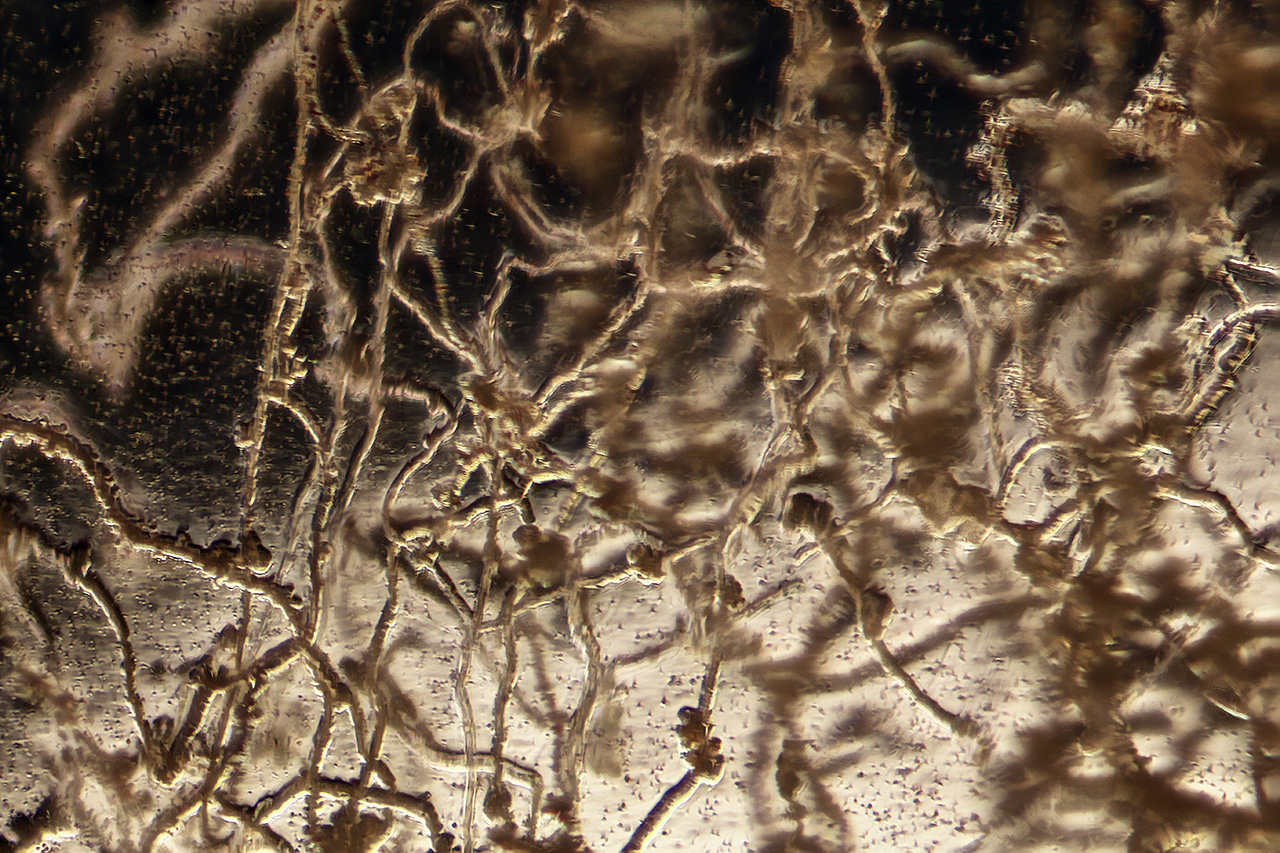
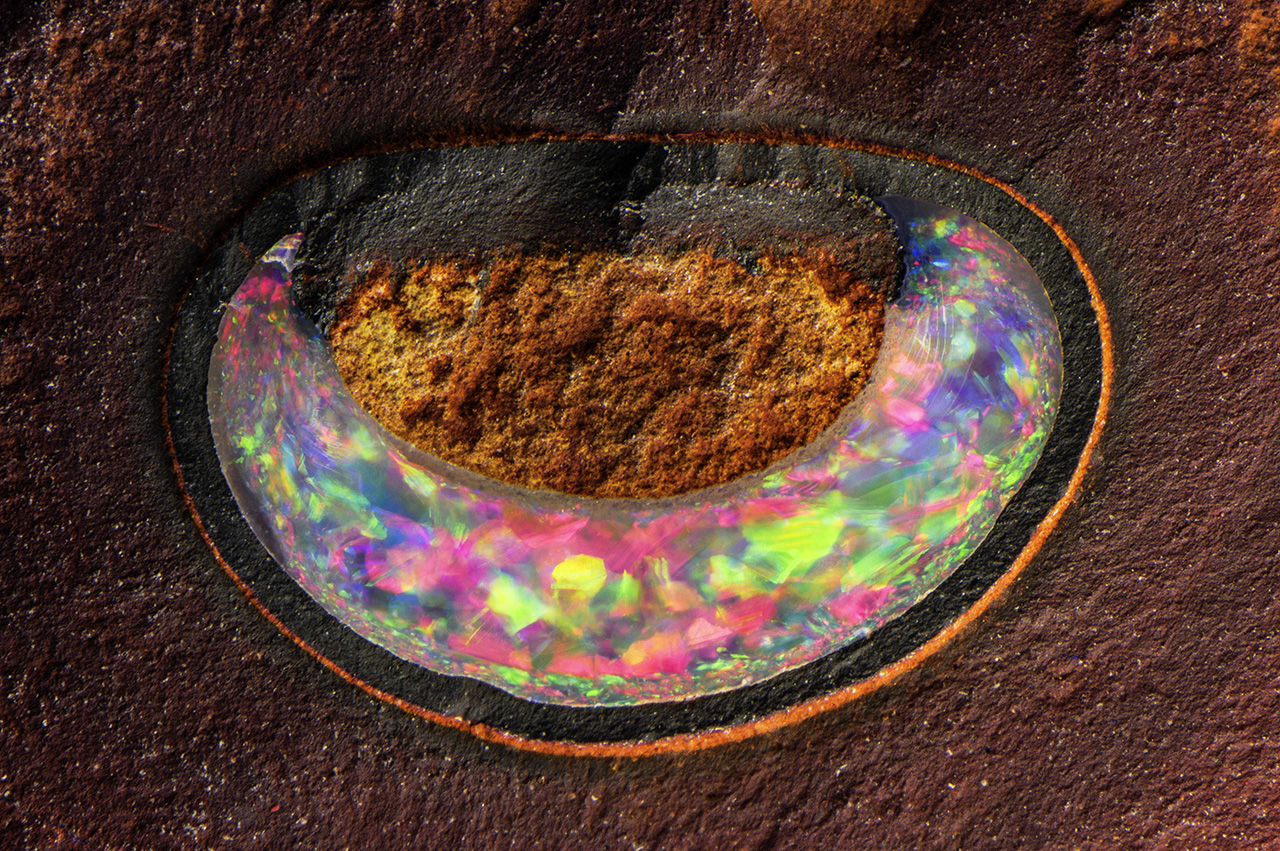
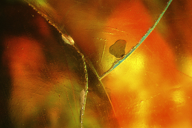
Some opals are prone to crazing or cracking, which can be hidden by filling the cracks with oil or resin. Trapped bubbles in the cracks of this opal reveal the treatment. Field of view 2.15 mm.
When examined under the microscope, certain visual features, such as those shown here, are good indications that an opal has been treated to improve its appearance or durability.

One of the more prominent opal imitations was introduced in the early 1970s by John Slocum of Rochester, Michigan. In this imitation, thin sheets of metal foil were embedded in glass to produce thin-film interference colors resembling “play-of-color” in natural opal. In 1972, Pierre Gilson began to manufacture a true synthetic opal (Gübelin and Koivula, 2005) that did not require polymer impregnation. Synthetic and imitation opals have been produced over the years by other manufacturers including Kyocera, Almaztechnocrystal, and Openallday Pty. Ltd, but the most recent development is an opal-like plastic manufactured by Kyocera and others in Japan (Renfro and Shigley, 2017). This new material is 80% plastic and 20% silica. It can be produced in large sizes, giving rise to industrial applications such as use in eyeglass frames and watches.

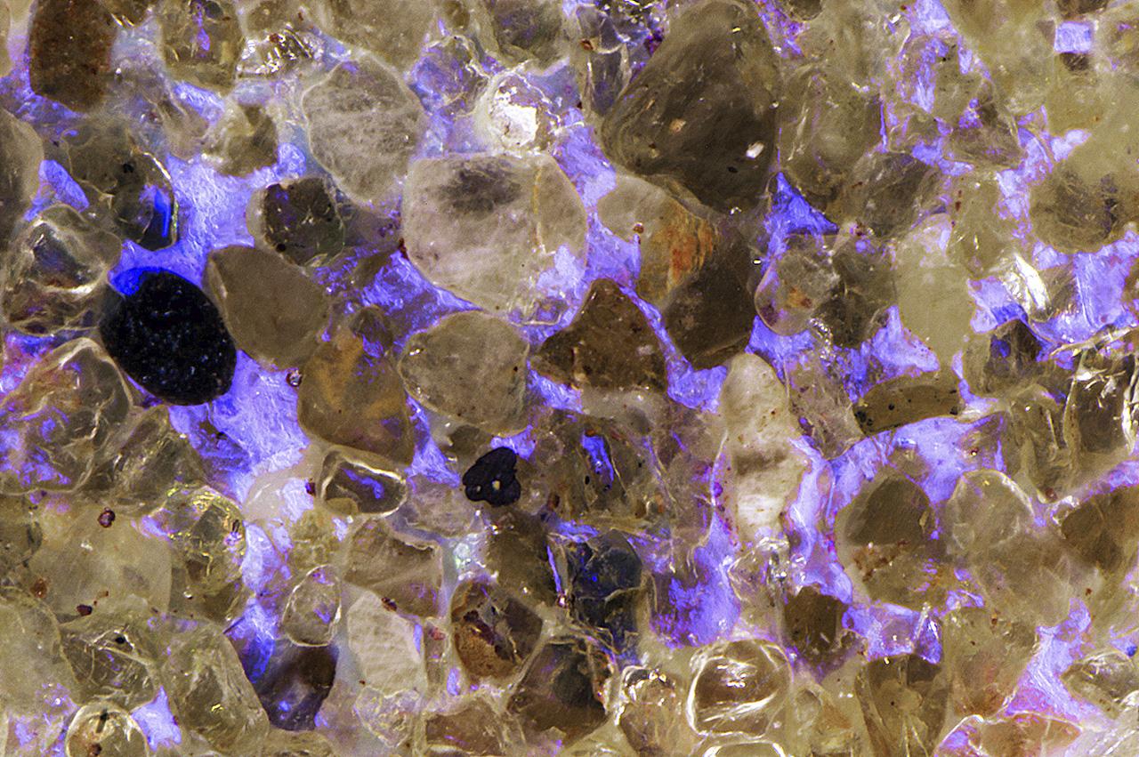
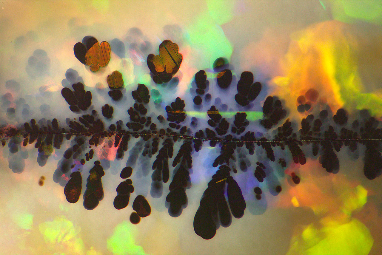
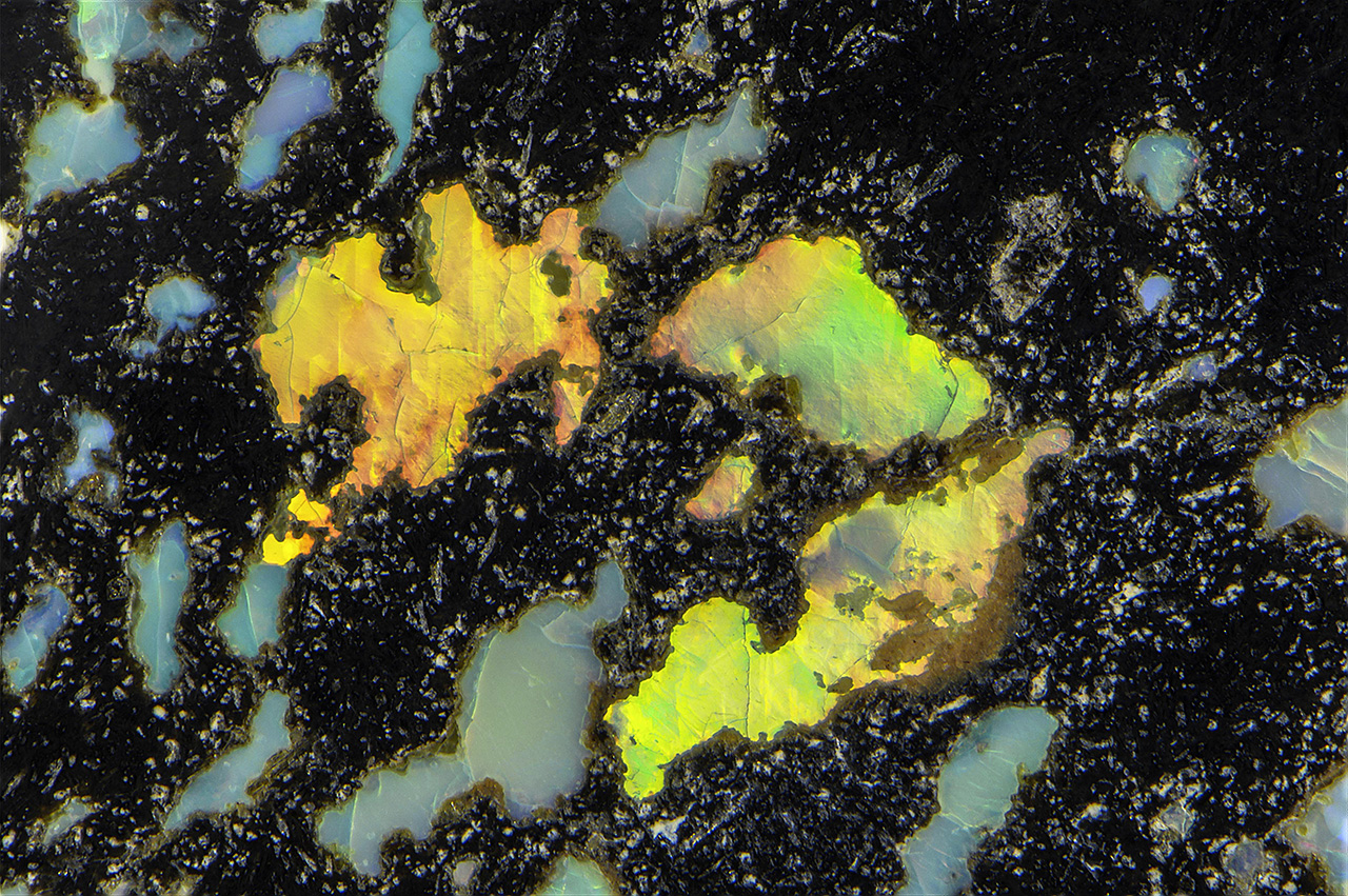
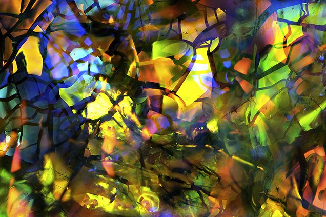
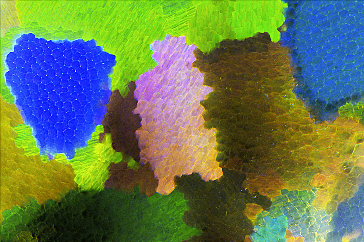
Slocum Stone opal imitation glass was manufactured by John Slocum of Michigan in the early 1970s. Thin layers of metallic film fragments suspended in the glass matrix are responsible for the interference colors observed. Field of view 5.75 mm.
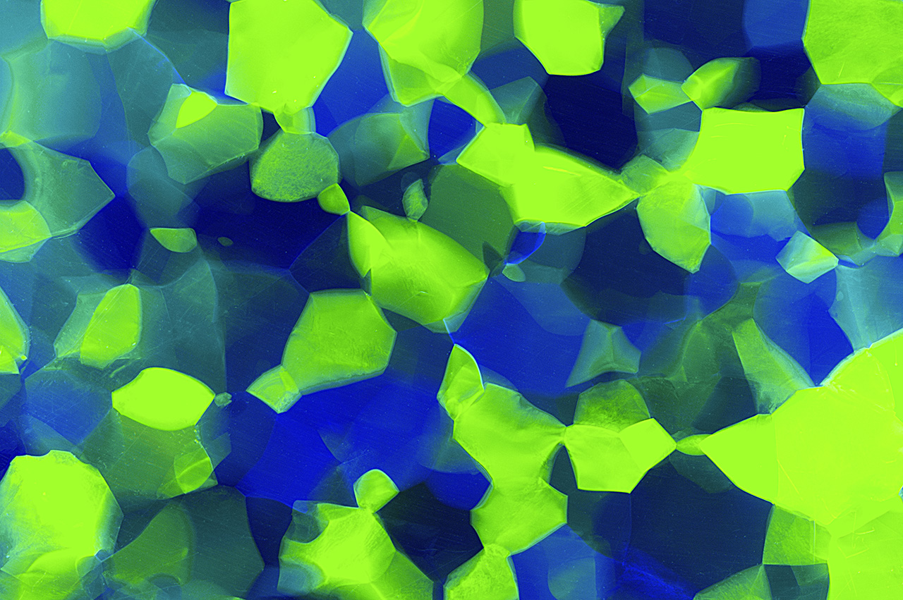
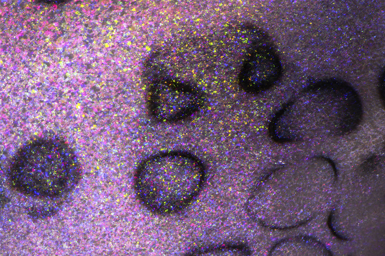
Synthetic and imitation opals commonly show features resulting from their formation that differ in appearance from those seen in natural opals.

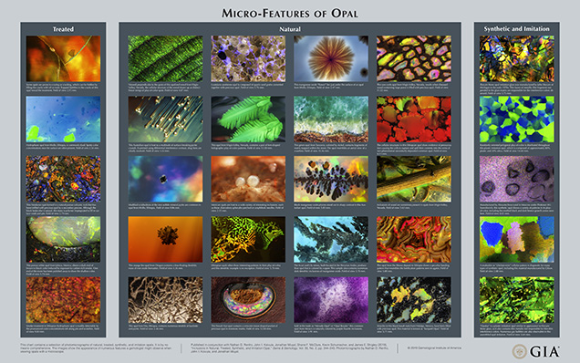
While the images in the accompanying wall chart are by no means comprehensive, they do represent a wide variety of the micro-features one might encounter in natural, treated, synthetic, and imitation opals.
Micro-Features of Opal
This chart contains a selection of photomicrographs of natural, treated, synthetic, and imitation opals. It is by no means comprehensive. The images show the appearance of numerous features a gemologist might observe when viewing opals with a microscope.

Mr. Renfro is manager of colored stones identification, and John Koivula is analytical microscopist at GIA in Carlsbad, California. Mr. Muyal is a staff gemologist, Mr. McClure is global director of colored stone services, Mr. Schumacher is a photo and video producer for Gems & Gemology, and Dr. Shigley is distinguished research fellow at GIA in Carlsbad.

Gübelin E.J., Koivula J.I. (2005) Photoatlas of Inclusions in Gemstones, Vol. 2, Opinio Verlag, Basel, Switzerland, p. 486. 

Renfro N.D., McClure S.F. (2011) Dyed purple hydrophane opal. G&G, Vol. 47, No. 4, pp. 260–270, http://dx.doi.org/10.5741/GEMS.47.4.260

Renfro N.D., Shigley J.E. (2018) Lab Notes: New plastic opal imitation from Kyocera. G&G, Vol. 54, No. 1, pp. 60–62.

Renfro N.D., York P. (2011) Lab Notes: Opal: Clarity-enhanced, with artificial matrix. G&G, Vol. 47, No. 4, pp. 312–313.

Rondeau B., Fritsch E., Mazzero F., Gauthier J.-P., Cenki-Tok B., Bekele E., Gaillou E. (2010) Play-of-color opal from Wegal Tena, Wollo Province, Ethiopia. G&G, Vol. 46, No. 2, pp. 90–105, http://dx.doi.org/10.5741/GEMS.46.2.90

Williams B., Williams C. (2011) Smoke treatment in Wollo opal. http://stonegrouplabs.com/wp/wp-content/uploads/2013/01/SmokeTreatmentinWolloOpal.pdf [date accessed: June 24, 2019].