Gemological and Chemical Characteristics of Natural Freshwater Pearls from the Mississippi River System

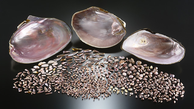
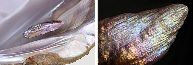
The Carlsbad laboratory received 854 natural freshwater pearls from Kari Anderson (Kari Pearls, Muscatine, Iowa), who stated that they were collected from the Mississippi River system (figure 1). Although the time and location of recovery and the mollusk species were not recorded, we were informed that all were recovered as a byproduct of the shelling business in the past couple of years. The samples ranged from 0.013 to 3.59 ct and measured from 1.63 × 1.19 mm to 9.87 × 8.52 × 5.89 mm. The shapes varied from baroque (the majority) to near-round, button, and oval. Many of the baroque pearls exhibited the unique “wing” or “feather” form (Anderson mentioned that the divers refer to these as “spike” pearls) typically associated with American natural freshwater pearls (J.L. Sweaney and J.R. Latendresse, “Freshwater pearls of North America,” Fall 1984 G&G, pp. 125–140). The wing pearls are elongated, with a roughly triangular shape, and usually taper to more pointed ends. Most possessed an uneven rippled surface along their lengths. The surface texture and shape of the wing pearls are identical to the lateral “teeth” areas found on the mussel shells that host these organic gems (figure 2, left).

The pearl colors also varied widely in hue, tone, and saturation. The range of hues included very light pink, purplish pink, orangy pink, brownish orange, and brownish purple to brown. Many displayed a pronounced orient effect (iridescence or multiple colors on or below the surfaces), creating a beautiful shimmering appearance (figure 2, right). Moreover, some exhibited high metallic-like surface reflections that resulted in excellent luster. Classifying the colors was often quite complex, as the overtone and orient were strong enough to affect the actual bodycolor and the combinations produced different colors when viewed from different angles. Experience has shown that in most cases a pearl’s color is directly related to the mussel species and the color of the shell’s interior, though water environment and the nutrients available during formation also play an important role. The heelsplitter (Potamilus alatus), purple wartyback or purple pimpleback (Cyclonaias tuberculate), and bankclimber (Plectomerus dombeyanus) mussel species shown in figure 1 possess pink and purple interior surfaces. All have been known to produce pearls with desirable color.
The lightly colored samples exhibited moderate to strong yellow or bluish yellow fluorescence when exposed to long-wave ultraviolet radiation (365 nm), while the darker samples exhibited the same colors with weaker intensities. X-ray fluorescence imaging was used to help check the growth environment. The majority of the samples fluoresced weak to strong greenish yellow due to manganese content, thus confirming their freshwater origin. As with the long-wave UV reactions, the darker samples displayed weaker reactions, and this fluorescence quenching is related to the concentration of coloring pigments present (H. Hänni et al., “X-ray luminescence, a valuable test in pearl identification,” Journal of Gemmology, Vol. 29, No. 5/6, 2005, pp. 325–329). A few of the samples also exhibited a moderate to strong orangy reaction in some surface areas.
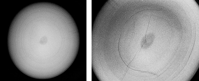
One hundred samples encompassing a broad range of colors, shapes, surface qualities, and sizes were selected in order to study their internal structures and trace element concentrations using real-time microradiography (RTX) and laser ablation–inductively coupled plasma–mass spectrometry (LA-ICP-MS), respectively. The baroque pearls showed growth structures that followed their shapes. The number and visibility of these structures varied from pearl to pearl. In certain orientations the growth structures resembled “linear features” in some wing pearls. Dark organic-rich and void centers were observed in some of the symmetrically shaped samples (i.e., button and oval). Although void structures could be considered characteristic of non-bead cultured (NBC) pearls, the voids in these samples appeared ovoid and relatively small (e.g., figure 3) and unlike the “twisted” void-like structures typically encountered within freshwater NBC pearls. Chemical compositions were analyzed using the same parameters published in a recent study (A. Homkrajae et al., “Provenance discrimination of freshwater pearls by LA-ICP-MS and linear discriminant analysis (LDA),” Spring 2019 G&G, pp. 47–60). Three ablation spots were tested on each sample, and results for the 22 elements selected are shown in table 1. The majority contained high Mn content, as expected for freshwater pearls and corresponding with the X-ray fluorescence results seen from the majority. Only one sample showed Mn levels below 100 ppmw in two spots at 89.7 and 64.2 ppmw, which is unusual for this pearl type. Yet both spots matched positions for freshwater pearls in the ternary diagram of Ba, Mg, and Mn (see Homkrajae et al., 2019), indicating a likely freshwater origin. Chemical data for the analyzed spots, especially the four discriminator elements Na, Mn, Sr, and Ba (bolded in table 1), showed results comparable with those of the American natural (USA-NAT) pearls group previously reported. The majority of the analyzed spots fell within the same region as the USA-NAT group in the Sr and Na content plot (figure 4). Furthermore, the linear discriminant analysis (LDA) method developed during the previous study discriminated 70% of these samples as USA-NAT.


Pearls exhibiting six different colors—pink, purplish pink, orangy pink, pinkish purple, brownish orange, and orangy brown—were selected in order to study their spectroscopic characteristics. Apart from the brownish orange sample, each possessed orient. The analysis was conducted on the most homogeneously colored area of each sample. Raman spectroscopic analysis with a 514 nm argon-ion laser revealed aragonite peaks at 701, 703, and 1085 cm–1 along with strong polyene peaks between the approximate ranges 1119–1126 and 1503–1508 cm–1. UV-Vis reflectance spectra were recorded from 250 to 800 nm, and each spectrum revealed a clear absorption feature at about 280 nm, possibly associated with the protein conchiolin and usually observed in nacreous pearls (figure 5). The reflectance patterns of the pearls are associated with the bodycolors, and the reflectance levels decreased as the sample color became more saturated. The samples with a pink component showed reflection minima in the blue to yellow range corresponding to their bodycolors, with a reflection minimum centered around 506 nm. The results corresponded with previous findings on natural-color freshwater cultured pearls from Hyriopsis species (S. Karampelas et al., “Role of polyenes in the coloration of cultured freshwater pearls,” European Journal of Mineralogy, Vol. 21, No. 1, 2009, pp. 85–97; A. Abduriyim, “Cultured pearls from Lake Kasumigaura: Production and gemological characteristics,” Summer 2018 G&G, pp. 166–183). Surface observations and spectroscopic studies did not reveal any sign of color treatment, which indicates the pearls’ natural color origin.

The Mississippi River and its tributaries are renowned for the diversity of native American mussels found in their waters. The extensive variety of species from the Unionidae family continues to produce a variety of colored natural pearls. This was a great opportunity to study and document the gemological and chemical characteristics of these unique freshwater natural pearls. The data collected enlarges GIA’s pearl identification database and will provide useful reference material in the years to come.


