Green-Blue Maxixe-Type Beryl
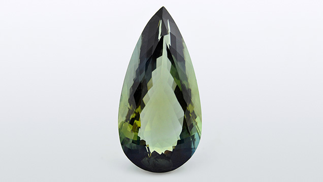
Recently, a transparent greenish blue pear-shaped mixed-cut sample (figure 1) was submitted for identification to the Gem Testing Laboratory in Jaipur. The 54.21 ct specimen (40.00 × 20.14 × 12.63 mm) was relatively clean to the unaided eye. Its refractive index (RI) of 1.582–1.590, birefringence of 0.008 with a uniaxial negative optic sign, and hydrostatic specific gravity (SG) of 2.71 suggested a beryl, which was later confirmed with Fourier-transform infrared (FTIR) and Raman spectroscopy. The specimen’s natural origin was established by zones of fine growth tubes, planes of platelets (usually ilmenite, although not identified here) oriented along the basal plane, and birefringent crystals.
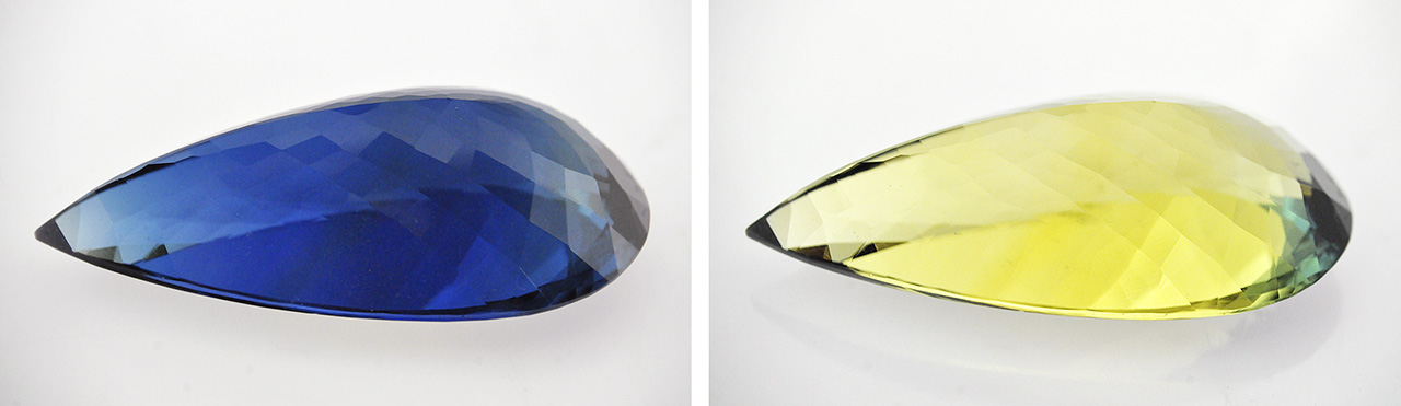
Identification of the stone as beryl was straightforward, but its unusual color and striking dichroism invited further study. It displayed deep blue and yellow as the two principal colors (figure 2); the deep saturated blue resembled the color of top-quality sapphires. Such strong dichroism was reminiscent of Maxixe-type beryls, although their dichroic colors are usually deep blue and colorless. Careful examination of the specimen showed a deep saturated blue o-ray and a yellow e-ray. Maxixe-type beryls are known to display deep blue color absorption along the o-ray direction (R. Webster, Gems, 5th ed., Butterworth-Heinemann, London, 1994, pp. 124–127) and colorless along the e-ray direction. An opposite pattern of deep blue (e-ray) and pale greenish blue (o-ray) absorptions was reported previously in an aquamarine by this author (Fall 2014 GNI, pp. 244–245).

of bands between 500 and 700 nm (o-ray) that are associated with radiation-induced color
centers.
Further analysis with UV-Vis-NIR spectroscopy confirmed the cause of color. Polarized spectra (figure 3) revealed a series of bands between 500 and 700 nm along the o-ray direction, but only a broad absorption feature at ~690 nm along the e-ray; these features are typically associated with the radiation-induced color centers observed in Maxixe-type beryl (see I. Adamo et al., “Aquamarine, Maxixe-type beryl, and hydrothermal synthetic blue beryl: Analysis and identification,” Fall 2008 G&G, pp. 214–226).
Gemological properties along with absorption spectra and the pleochroic color directions were sufficient to establish the identity of this submitted specimen as Maxixe-type beryl. This was the first time we had encountered a green-blue Maxixe-type beryl, although there exists a previous report of such a beryl (see K. Nassau et al., “The deep blue Maxixe-type color center in beryl,” American Mineralogist, Vol. 61, 1976, pp. 100–107). On the basis of pleochroic colors, it can be deduced that the greenish face-up appearance of this specimen was due to the overlap of blue and yellow color components. The supplier of the specimen informed us that the specimen is reportedly from Mozambique.


