Natural Blisters with Partially Filled Areas

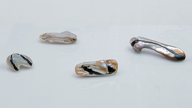
Natural blisters and blister pearls have been the subject of previous reports in G&G (see Lab Notes from Fall 1992, Spring 1995, Winter 1996, and Winter 2015, and Gem News International from Fall 2001 and Winter 2009). In February 2016, four large “pearls” (figure 1) were submitted to GIA’s Bangkok laboratory for identification. On first impression they appeared to differ from most pearls or blister pearls examined in the past. The specimens ranged from approximately 25.06 × 18.31 × 13.41 mm to 55.90 × 13.89 × 7.96 mm, and they weighed 32.33, 37.20, 41.41, and 52.17 ct. Two of the items were white, and the other two were silver and orangy brown.
All the samples had eye-visible areas on their bases and around their outlines where they had obviously been worked or cut to either remove them from their shell hosts or improve the symmetry (in some cases both). These are telltale signs of blisters and blister pearls, since both must be removed from the shell to be presented in loose form. What caught our attention was the fact that all four items possessed dark or cream bands on their bases (figure 2). These bands appeared to be organic-rich formations, noted in some pearls and more commonly in shells, yet this did not turn out to be the case in three of the samples.

The items were considered blisters rather than blister pearls (E. Strack, Pearls, Ruhle-Diebener-Verlag, Stuttgart, Germany, 2006, pp. 115–127). This determination was based on external appearance and features, the “work” that had taken place on them in relation to where they were likely removed from the shells, and the results of real-time X-ray microradiography (RTX), which revealed growth arcs following the shape of the blisters to varying degrees.
The curving black band on the base of the smallest white blister contained translucent to opaque organic-looking material characteristic of conchiolin (figure 3A), one of the constituents of pearls and shells. The remaining three blisters had structures within their bands that did not match the structure observed in the first blister. The bands in the two orangy brown blisters consisted of an essentially transparent near-colorless substance in which minute black pinpoint particles imparted an overall black color (figure 3B). Meanwhile, the band in the remaining white blister showed areas of completely transparent near-colorless material and other areas of the same near-colorless material, mixed with small pieces of what appeared to be shell fragments. Distorted bubbles were clearly visible in the transparent areas on the base of the partially filled white blister (figure 3C) and one of the colored blisters (figure 3D); no obvious bubbles were seen in the other blister. RTX also revealed the extent of the filling on the bases of the three blisters.

Raman spectroscopy of the near-colorless filled areas of the two colored blisters did not show any polymer or resin peaks that matched those found in the white blister’s filling. Therefore, we conducted basic testing on all three samples with a very carefully placed hot point in areas of the filling where some damage or abrasion already existed. The unmistakable plastic odor and melting of the tested areas was enough to confirm the artificial nature of the fillers. Interestingly enough, the fillers did not display a noticeable fluorescence under long-wave or short-wave ultraviolet light, but the two orangy brown blisters did exhibit distinct orange to orange-red fluorescence, which is characteristic of the porphyrins (naturally occurring pigments) known to exist in Pteria species shells of similar coloration (L. Kiefert et al., “Cultured pearls from the Gulf of California, Mexico,” Spring 2004 G&G, pp. 26–38). Out of curiosity, we also checked the dark conchiolin-rich band in the smaller white blister with the hot point and fluorescence. It was no surprise to smell a distinctly unpleasant organic reaction from the band and see a weak chalky yellowish reaction under UV lighting.
These four blisters were good examples of this material, and three of them were the first partially filled blisters to be examined by GIA’s Bangkok laboratory. The three partially filled blisters show that even material with relatively low market value may be treated in some way, and buyers should always be aware of what is being offered to them.


