Multiphase Fluid Inclusions in Blue Sapphires from the Ilmen Mountains, Southern Urals
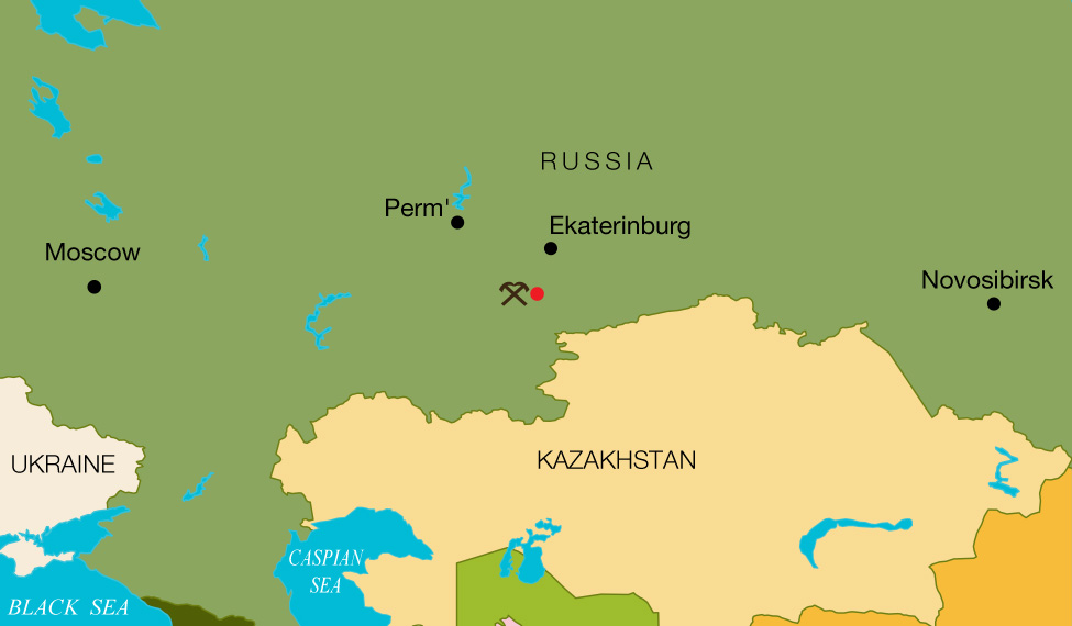
Most gem-quality sapphires are found in placers that originate from the weathering of primary deposits. Discovering a mineral in situ, or within the host rocks, is rare. In August 2015, some of the authors visited a corundum deposit in the Ilmen Mountains in the southern Urals of Russia (figure 1) to collect the stones in situ for research. This is a primary source where blue sapphire megacrysts are found exclusively in syenite pegmatites. The occurrence is located inside the Ilmen State Reserve, and its commercial exploitation is forbidden.
Corundum syenite pegmatites in the Ilmen Mountains present an REE-rich source of transparent to translucent blue sapphire crystals measuring up to 5.9 × 4.2 cm (figure 2). But the brittle deformation throughout the gem-bearing rocks, which appears to be linked to syntectonic and post-tectonic processes that occurred at the deposit and the surrounding area, increased the amount of fissures in the corundum. As a result, the sapphires are of a lower quality and can only be faceted into small stones. Six sapphire wafers and nine petrographic thin sections of mineral from corundum-bearing rocks of this occurrence were recently examined at GIA’s Carlsbad lab.

Laser ablation–inductively couple plasma–mass spectrometry (LA-ICP-MS) and electron microprobe analysis (EPMA) indicated medium-rich Fe (2470–3620 ppmw), high Ga (190–280 ppmw), and low Mg (3–9 ppmw) with Ga/Mg 29, all in the range of magmatic sapphires (see J.J. Peucat et al., “Ga/Mg ratio as a new geochemical tool to differentiate magmatic from metamorphic blue sapphires,” Lithos, Vol. 98, No. 1–4, 2007, pp. 261–274).
Ferrocolumbite, zircon, and alkali feldspar group minerals, identified by confocal micro-Raman spectroscopy, were observed as syngenetic inclusions. Epigenetic muscovite and exsolved needles (most likely ilmenite) were found as well; along with syngenetic inclusions, they are common phases in mineral assemblages of Ilmen corundum syenite pegmatites (see V. F. Zhdanov et al., “The mineralogy of corundum pegmatite pit no. 298 of the Ilmen State Reserve,” Materialy k mineralogii Yuzhnogo Urala [Materials to Mineralogy of South Urals], AS USSR, 1978, pp. 92–97, in Russian). Wafers 200–400 µm in width were prepared in order to study fluid inclusions (FI) trapped within the sapphire crystals.
Using Raman spectroscopy, solid phases in needle and thin-film form were identified as diaspore (figure 3); both vapor and liquid phases were found to be CO2, with a Fermi doublet for the liquid phase at 1282.9–1283.2 and 1387.2–1387.6 cm–1, accompanied by less intense symmetrical bands (“hot bands”; see N.B. Colthup et al., Introduction to Infrared and Raman Spectroscopy, 2nd ed., Academic Press, New York, 1975) at approximately 1264 and 1408 cm–1 and a small band at 1370 cm–1 due to 13CO2. Raman spectra in the 2000–4000 cm–1 range did not show H2O-, CH4-, or N2-related bands. The less intense apparent maxima at 418, 578, and 750 cm–1 belong to sapphire vibration modes. The distance between Fermi doublet peaks (Δ) ranged from 104.1 to 104.4 cm–1 (again, see figure 10), occurring at about 0.7 g/cm3 CO2 density (see X. Wang et al., “Raman spectroscopic measurements of CO2 density: Experimental calibration with high-pressure optical cell (HPOC) and fused silica capillary capsule (FSCC) with application to fluid inclusion observations,” Geochimica et Cosmochimica Acta, Vol. 75, 2011, pp. 4080–4093). The distance was linked to the pressure at approximately 1.8 kbar for the trapping of fluid inclusions within the sapphire matrix (CO2 P-T isochores, or lines of constant density). The CO2 homogenization temperature (the temperature at which the fluid inclusion becomes a single-phase liquid or vapor) occurred in the liquid phase and measured between 30.1° and 30.7°C (figure 4), while the corundum-diaspore equilibrium was estimated at 510°–530°C (matching the previous estimation by V.A. Simonov, Usloviya mineraloobrazovaniya v granitnykh pegmatitakh [Conditions of Mineral Formation in Non-Granitic Pegmatites], Nauka, Novosibirsk, USSR, 1981, in Russian). Based on the determined pressure (around 1.8 kbar), the temperature of stability for the corundum-orthoclase-H2O system was higher than 690°C (H.S. Yoder and H.P. Eugster, “Synthetic and natural muscovites,” Geochimica et Cosmochimica Acta, Vol. 8, 1955, pp. 225–280), likely linked to the multistage geological history of blue sapphire crystallization along the P-T path in the Ilmen Mountains.


Further study of these sapphires is needed to better understand the geological history of this locality, but the information obtained can provide valuable clues about the characterization of gem corundum from secondary placers.


