X-ray Computed Microtomography Applied to Pearls: Methodology, Advantages, and Limitations
X-ray computed microtomography reveals the internal features of pearls with great detail. This method is useful for identifying some of the natural or cultured pearls that are difficult to separate using traditional X-radiography. The long measurement time, the cost of the instrumentation, and the fact only one pearl at a time can be imaged are some of this method’s disadvantages.
DATA DEPOSITORY
Supplementary Videos and Images (see below)
Summer, 2010
Depository Item 1
Figures 1a, 1b, 1c
3D model of Beaded Saltwater Cultured Pearl
3D model of Beaded Saltwater Cultured Pearl
Videos showing slices taken in three directions through the 3D model of the beaded saltwater cultured pearl in figure 4 (sample GGL19). All sections show reconstruction artifacts due to rotation: fine circles in the horizontal section (a) and a blurry rotation axis in perpendicular sections (b and c).
Depository Item 2
Figures 2a-2f
3D model of Non-beaded Saltwater Cultured Pearl
3D model of Non-beaded Saltwater Cultured Pearl
Videos showing slices taken in three directions of the 3D model (with and without shadows) of the non-beaded saltwater cultured pearl in figure 5 (sample SK-51). In the videos b, d, and f, structures produced by the grafted tissue have an inverted color; that is, these structures appear dark in the original videos (a, c, and e) instead of bright. All sections show reconstruction artifacts due to rotation.
Depository Item 3
Figures 3a-3c
3D model of Freshwater Natural Pearl
3D model of Freshwater Natural Pearl
Videos showing slices in three directions of the 3D model of the freshwater natural pearl in figure 6 (sample GGL03). All sections show reconstruction artifacts due to rotation.
Depository Item 4
Figures 4a-4c
3D Model of Saltwater Natural Pearl
3D Model of Saltwater Natural Pearl
Videos showing slices in three directions of the 3D model of the saltwater natural pearl (sample SK-46) presented in the figure 7. All sections show reconstruction artifacts due to rotation.
Depository Item 5

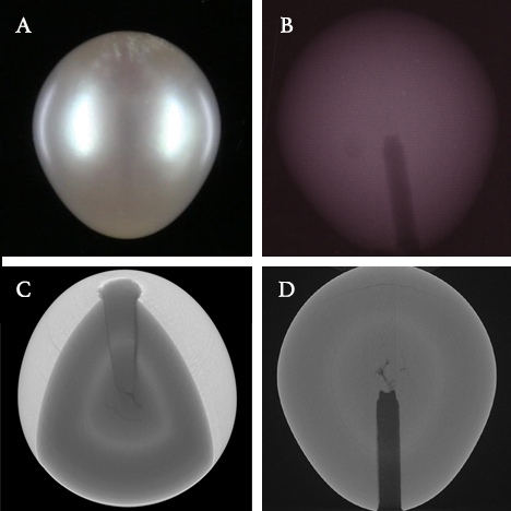
In this (a) gray-black half-drilled, drop-shaped, non-beaded freshwater cultured pearl
from Hyriopsis spp. (sample SK-54), a characteristic comma-shaped structure caused
by the grafted tissue was only visible in one direction in the radiographs (b). Tissue-related
structures (i.e., undulating growth lines) are clearly visible only in the μ-CT images (c, d).
from Hyriopsis spp. (sample SK-54), a characteristic comma-shaped structure caused
by the grafted tissue was only visible in one direction in the radiographs (b). Tissue-related
structures (i.e., undulating growth lines) are clearly visible only in the μ-CT images (c, d).
Figures 5e-5j
3D Model of Non-beaded Freshwater Cultured Pearl
3D Model of Non-beaded Freshwater Cultured Pearl
The videos show slices in three directions of the 3D model of this sample. In the videos b, d, and f, the tissue-related structures are shown with inverted colors (they appear bright). All sections show reconstruction artifacts due to rotation.
Depository Item 6

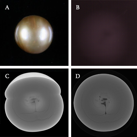
In this near-round, non-beaded freshwater cultured pearl (a) from Hyriopsis spp.
(sample GGL26), a mixture of structures representative of a cultured pearl are observed
in the radiograph (b). The μ-CT images (c, d) suggest that these structures are related
to more than one piece of grafted tissue.
(sample GGL26), a mixture of structures representative of a cultured pearl are observed
in the radiograph (b). The μ-CT images (c, d) suggest that these structures are related
to more than one piece of grafted tissue.
Figures 6e-6j
3D Model of Non-beaded Freshwater Cultured Pearl
3D Model of Non-beaded Freshwater Cultured Pearl
This is more evident in two of the videos (f and j), where the tissue-related structures are shown with inverted colors (they appear bright). Photo by A. Spingardi. All sections show reconstruction artifacts due to rotation.
Depository Item 7

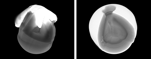
These 3D μ-CT images of a half-drilled, drop-shaped, saltwater natural pearl from Pteria spp. (sample SK-61) show the pearl mounted (left) and unmounted (right). The metal produces artifacts that partially mask the internal features; the structures indicative of a natural origin are much more visible in the unmounted pearl.


