An Unusual Treated Agate Presented as “Shi Zi Hong” Agate from Liangshan
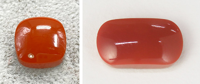
In October 2020, an orange-red cabochon sample was presented to the National Gold-Silver Gem & Jewelry Quality Supervision & Inspection Center (Sichuan) by a client who claimed it was a new type of Shi Zi Hong agate. The trader referred to it as “Meng Liao” agate (figure 1, left).
The sample weighed 5.74 ct, and the appearance was similar to Shi Zi Hong agate (example shown in figure 1, right). The spot RI reading was around 1.54, and the sample was inert to UV radiation. Microscopic examination showed cryptocrystalline texture with red pinpoint-like inclusions scattered throughout, most of which were sufficiently fine to appear as hazy clouds under 40× magnification.
Raman spectra of the substrate and red pinpoints were obtained using 532 nm laser excitation. According to the RRUFF online database, peaks at 126, 205, 261, 353, and 464 cm–1 indicated that the main constituent of the substrate was quartz, while the red pinpoints matched that of hematite, with peaks at 224, 244, 291, 410, 610, and 1320 cm–1 (figure 2). Both were consistent with Shi Zi Hong agate. A sharp peak at 501 cm–1, seldom observed in Shi Zi Hong agate, appeared in the spectra of the sample. This significant peak was assigned to stretching vibrations of (SiO4)4– in the moganite structure.

Figure 2. Under magnification and using fiber-optic illumination, some fine pinpoints were distributed as hazy clouds in the rough agate. Raman spectroscopy identified the main constituent of substrate in the sample as quartz (green trace) and red pinpoint as hematite (red trace), both consistent with “Shi Zi Hong” agate (indicated by the purple trace and the black trace). The sample also showed a sharp peak at 501 cm–1 and a broad band at 660 cm–1, neither of which are seen in genuine “Shi Zi Hong” agate. Photomicrograph by Su Xu; field of view 0.22 mm. Spectra offset for clarity.
Based on a negative correlation between the relative content of moganite and the crystallinity of agate (Zhou Dan-yi et al., “Study on the relationship between the relative content of moganite and the crystallinity of quartzite jade by Raman scattering spectroscopy, infrared absorption spectroscopy and X-ray diffraction techniques,” Rock and Mineral Analysis, Vol. 3-4, No. 6, 2016, pp. 652–658), we concluded that the crystallinity of natural Shi Zi Hong agate was much higher than that of the sample. Another obvious anomaly was the broad band at 660 cm–1, which was found in the spectrum of the sample but was seldom observed in Shi Zi Hong agate, and this deserved further investigation.The client admitted that the “Meng Liao” agate was actually a normal brownish yellow agate heated by a low-temperature process. Meanwhile, he loaned us an untreated rough stone (figure 3) for Raman analysis of its body and very fine yellow pinpoint inclusions.

Figure 3. The untreated brownish yellow rough stone, approximately 17.0 × 22.0 × 1.5 mm, loaned by the client for analysis. Photo by Xiaoping Shi.
The substrate of the untreated rough stone and the “Meng Liao” agate sample submitted to the lab showed almost the same peaks in the region of 1000–100 cm–1, suggesting that they might be homologous with each other. The spectrum of the yellow pinpoint, exhibiting peaks at 299, 389, 415, and 550 cm–1 and lacking a 660 cm–1 band, matched the Raman spectrum for goethite and was related to the formation of color in the rough, as shown in figure 4. According to a previous report (D.L.A.de Faria and F.N. Lopes, “Heated goethite and natural hematite: Can Raman spectroscopy be used to differentiate them?” Vibrational Spectroscopy, Vol. 45, No. 2, 2007, pp. 117–121), the disordered hematite structure was initially formed by dehydration and caused the relative intense band at 660 cm–1 by heating goethite at a low temperature (~140°C to 360°C).
Figure 4. Raman spectra of the brownish yellow rough stone: Peaks at 299, 389, 415, and 550 cm–1 indicated goethite. Peaks at 126, 205, 261, 353, 464, and 501 cm–1 were consistent with the treated agate sample submitted to the lab. Spectra are offset for clarity.
The natural hematite in Shi Zi Hong agate, by contrast, has features including a very weak 660 cm–1 band and a high degree of order, by which genuine Shi Zi Hong agate can be distinguished from the brownish yellow agate that acquired an orange-red color by artificially heating goethite. These results show that the agate submitted by the trader as “Meng Liao” agate, resembling Shi Zi Hong agate, can be readily detected by its unique Raman spectrum displaying an obvious broad band at 660 cm–1 and a sharp peak at 501 cm–1. Consumers in the market for Shi Zi Hong agate should watch out for this treated material.

