Cummingtonite Needles Encased by Quartz in a Rhodonite
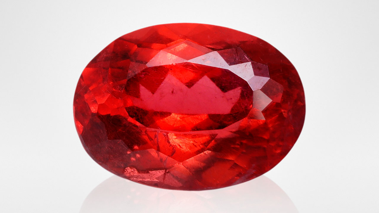
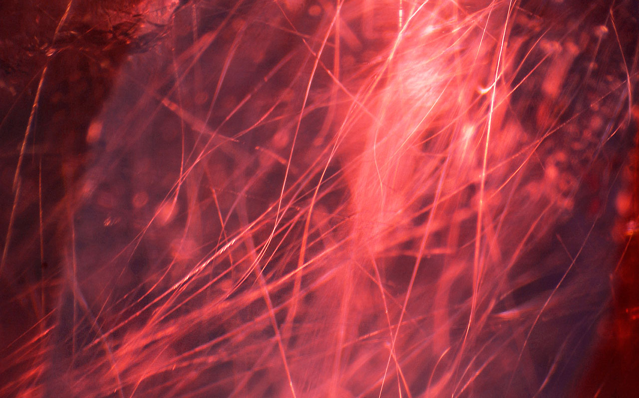
Recently, the authors examined a 7.38 ct gem-quality rhodonite exhibiting a vivid orangy red color and good transparency (figure 1). Gemological testing, Fourier-transform infrared spectroscopy, and Raman spectroscopy identified it as rhodonite. This gemstone had a refractive index of 1.733–1.747 and specific gravity of 3.46. Most rhodonite is opaque and is used as an ornamental stone, and transparent facet-grade material is very rare. Microscopic observation revealed an abundance of long, curved needles scattered randomly throughout the gem host (figure 2), consistent with previous studies. Occasional fluid inclusions were also seen.
A previous publication suggests that these curved needles are cummingtonite, an amphibole group mineral with a chemical formula of (Mg, Fe2+)2(Mg,Fe2+)5Si8O22(OH)2 (P. Leverett et al., “Ca-Mg-Fe-rich rhodonite from the Morro da Mina mine, Conselheiro Lafaiete, Minas Gerais, Brazil,” Mineralogical Record, Vol. 39, 2008, pp. 125–130). Raman spectroscopy confirmed the identity of one needle as cummingtonite, displaying characteristic peaks at 190, 667, and 1036 cm–1 when the laser beam was focused on the center of it. However, several new peaks appeared when the laser beam moved to the boundary between the needle and the rhodonite, including a 208 cm–1 peak that matched with quartz. Such a finding is new to the authors, since needle inclusions encased in another mineral have rarely been reported. To further investigate this finding, more than 10 points were selected, and the results identified quartz between the cummingtonite needle and the rhodonite host. All the tested Raman spectra were compared to the Raman online database.


To further explore the relationship between rhodonite, quartz, and cummingtonite needles, a 3D Raman map was performed on a selected area of 3 × 14 × 12 μm with a step size of 2 μm. After running for 17.25 hours, more than 500 spectra were recorded. Three feature peaks were selected for image reconstruction: 3658 cm–1 for cummingtonite, 113 cm–1 for rhodonite, and 208 cm–1 for quartz (figure 3). Due to the similarity between the Raman spectra of cummingtonite and rhodonite, the main peaks were not chosen for mapping. The OH-related peak at 3658 cm–1 was only observed in cummingtonite, enabling us to distinguish the two minerals. The Raman mapping image is shown in figure 4, with blue indicating the host, green for quartz, and red for cummingtonite. These images revealed that the detection of quartz between rhodonite and cummingtonite was not by accident but indicated a considerable presence of quartz inclusions. Previous studies have reported quartz as an isolated and random mineral inclusion in rhodonite from Australia (e.g., P. Millsteed et al., “Inclusions in transparent gem rhodonite from Broken Hill, New South Wales, Australia,” Fall 2005 G&G, pp. 246–254). Based on our observation of the position of the quartz, we speculate that it may be the product of the decomposition of cummingtonite under certain temperatures and pressures.
This intricate inclusion association pattern may offer a new perspective on the origin of curved needles in rhodonite. This case also highlights the usefulness of Raman mapping as a tool for nondestructive analysis of inclusions.


