Grayish Blue CVD Diamond Colored by GR1 and SiV–

Silicon is a common impurity in chemical vapor deposition (CVD) laboratory-grown diamond. The silicon-related SiV– center, which has zero-phonon lines at 736.6 and 736.9 nm and is active in absorption and luminescence, is widely considered an identifying feature of CVD diamond. Generally, the SiV– center is detected by photoluminescence spectroscopy and is not observed using absorption spectroscopy for most CVD synthetics due to the low silicon content. The absorption spectra may reveal the presence of the SiV– center in the case of a relatively high silicon content, but rarely in amounts significant enough to affect the color. When the silicon content becomes particularly high, the absorption of SiV– is in sufficient concentration to influence the bodycolor of a diamond. It has been reported that the color of silicon-doped blue CVD diamonds produced by the PDC Company is attributed to strong absorption of the rather intense SiV– center (A. Peretti et al., “New generation of synthetic diamonds reaches the market (Part A): Identification of CVD-grown blue diamonds,” Contributions to Gemology, No. 14, 2013, pp. 3–20).

National Gemstone Testing Center’s (NGTC) Beijing laboratory recently received a 1.63 ct grayish blue princess cut (figure 1, left) for identification. Standard testing identified it as a CVD synthetic diamond. The sample showed an even color distribution. Microscopic investigation revealed abundant non-diamond carbon inclusions in the form of both crystals and clouds, with the clouds distributed along the growth layer (figure 1, right). The visible/near-infrared (Vis-NIR) spectrum (figure 2), recorded at liquid nitrogen temperature, included a strong broad SiV– center defect at 737 nm next to an equally strong GR1 center. The diamond’s grayish blue bodycolor was attributed to both the strong GR1 center and the high intensity of the SiV– center, which produced preferential absorption in the red part of the spectrum. In addition, weak absorptions at 830, 856, and 946 nm (the SiV0 center) were observed, which were also reported in CVD synthetic diamonds with high silicon content (Peretti et al., 2013; Z. Song et al., “Silicon-doped CVD synthetic diamond with photochromic effect,” Journal of Gems and Gemmology, Vol. 18, No. 1, 2016, pp. 1–5).
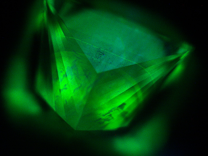

Infrared spectroscopy showed features typical for type IIa diamond (i.e., no defect-related absorptions were detected). The diamond was inert to short-wave and long-wave ultraviolet illumination. Under the ultra-short-wave UV radiation of the DiamondView, the sample fluoresced green, with minor areas showing some blue dislocations. It also showed subtle growth striations and two parallel planes fluorescing stronger than the bulk of the crystal (figure 3). The sample did not phosphoresce. Photoluminescence (PL) spectra taken with various laser excitation wavelengths and at liquid nitrogen temperature (figure 4) showed the presence of radiation-related features including very strong 3H at 503.5 nm, strong GR1 at 741 nm, and weak 486.3 nm. The PL spectra also indicated strong nitrogen-vacancy centers at 575 NV0 and 637 NV– nm and multi-nitrogen defects such as H3 and H2, combined with the absence of the unassigned 596/597 nm doublet that is normally seen in as-grown CVD synthetics, suggesting that it had undergone post-growth high-temperature annealing before irradiation.
The PL spectrum with 532 nm laser excitation also exhibited a rather large silicon doublet peak at 736.6/736.9 nm that dwarfs all other PL peaks, including the GR1 in the spectrum. The intensity ratio between the 736.6/736.9 nm PL peak and the diamond Raman peak was about 52. The PL spectrum reaffirmed that the 737 nm absorption peak of the ultraviolet/visible spectrum was the SiV– defect, and the silicon content of this diamond was so high that it produced strong absorption in the red range of visible light and affected the color of the diamond together with the GR1 center.


