Prominent Growth Planes Observed in HPHT-Processed CVD Laboratory-Grown Diamonds

The laboratory-grown diamond industry is ever evolving and improving. With state-of-the-art growth and treatment technology, high-quality CVD diamonds, in terms of clarity and color, are now commonly seen on the consumer market. Laboratory-grown diamonds are occasionally submitted for natural diamond grading services, and recently GIA’s Hong Kong laboratory received a total of 43 undisclosed CVD-grown diamonds treated by high pressure and high temperature (HPHT), ranging from 0.70 to 2.63 ct in weight, from the same client. Laboratory-Grown Diamond Reports were issued for these synthetics, which received E–I color grades and VVS2–I2 clarity grades.
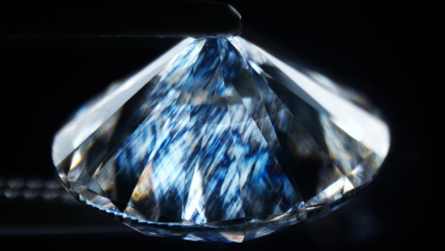
Four samples had a very eye-catching feature—a dark circle or ring at the center when viewed face-up without magnification. The ring appeared to be a thick grayish layer at the culet when viewed from the pavilion (figure 1). As the extraordinary feature looked shiny and resembled the separation plane often observed in assembled stones, questions about the true identity of these samples were raised.
Microscopic examination of these four samples revealed some interesting findings. There was a textured plane almost parallel to the table facet, each with a different appearance (figure 2): fine clouds all the way across; ripple-like waves with some “holes” (colorless areas); a mixture of both; and a ripple-like plane that was not visible all the way around.

An additional 26 samples had visible grayish/brownish planes either at the pavilion, which were much less prominent, or at the pavilion viewing through the table. Eight of these additional samples had a plane at an angle to the table facet. The plane appeared as a line with no color difference when immersed in water and viewed in a parallel direction to the growth plane. All 43 samples generally showed a banded and cross-hatched strain pattern when viewed with crossed polarizers (figure 3).

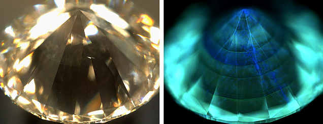
Spectroscopic analysis of the 43 samples yielded very similar results. They were either type IIa or type IIb and had a very strong silicon vacancy doublet peak (736.5 and 736.9 nm) in their photoluminescence spectra, typical of CVD-grown diamonds. PL spectra were also collected at the table and at the pavilion somewhere between the growth plane and the culet for the samples with a very prominent growth plane, but the data did not show any critical differences. The most diagnostic test was DiamondView imaging, as all 43 displayed very distinct growth layers (figure 4). It also revealed the HPHT treatment history that the CVD diamonds had experienced.

The uniformity of the growth layers observed in DiamondView imaging and under crossed polarizers, together with the immersion test, showed no evidence of assembly and therefore excluded the possibility of a doublet. The color making the growth plane visible is believed to come from the non-diamond carbon deposited between two growths. It could not be removed by the post-growth HPHT treatment.


