Bicolor Rough Diamond Crystals

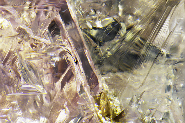
The GIA laboratory in Antwerp regularly receives pink diamond crystals for examination as part of the Diamond Origin Report. This recent service has allowed GIA researchers to study a greater number of rough diamonds in addition to their faceted counterparts. Two crystals, weighing 1.75 and 1.44 ct and both reportedly from Australia, were among those submitted. These were considered quite interesting, as they contained colorless and pink sections with distinct boundaries (figure 1).
The color in the vast majority of naturally pink diamonds is attributed to a broad absorption band at about 550 nm within the visible absorption spectrum. This band generally results from distortion of the crystal lattice from plastic deformation due to stress after crystal growth. However, much remains unknown about the actual formation and configuration of this feature. Therefore, these two crystals provide a unique chance to study natural diamond formation and the origin of pink color in greater detail.
The pink section of both analyzed samples likely experienced great stress in order to undergo the plastic deformation necessary to impart the pink color. The colorless sections were not similarly deformed so presumably they represent a different and later growth event (figure 2).

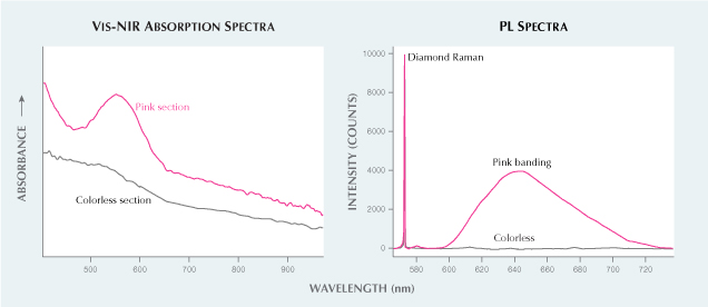
Visible-NIR absorption spectra collected from the pink and near-colorless sections of the diamonds show differences in the 550 nm region. Observed in the spectrum for the pink portion is the expected absorption at 550 nm, while the spectrum for the colorless portion shows a lack of absorption (figure 3, left).

For the 1.44 ct crystal, we also collected infrared (IR) absorption spectra and photoluminescence (PL) spectra from both the pink and the colorless sections using 455 and 532 nm laser excitation wavelengths. The IR absorption spectra for both sections indicate type Ia diamonds with saturated concentrations of nitrogen. Due to this saturation, it was not possible to determine the total nitrogen content or the ratio of the A and B nitrogen centers, which could have helped in further distinguishing these two sections.
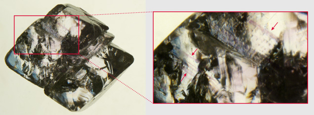
Within the PL spectra, the peak widths for the diamond Raman peak and the H3 peak were comparable in both the pink and colorless sections. Additionally, a PL peak at 676 nm was detected in both sections; although its configuration is unknown, it was documented previously in other rough pink diamonds (E. Gaillou et al., “Spectroscopic and microscopic characterizations of color lamellae in natural pink diamonds,” Diamond and Related Materials, Vol. 19, No. 10, 2010, pp. 1207–1220). The other notable feature detected by PL mapping was a broad (~600–730 nm) emission band (figure 3, right) that coincided with the pink banding revealed by immersion in methylene iodide (figure 4). Its detection is consistent with other pink diamonds and likely related to the 550 nm absorption band (e.g., S. Eaton-Magaña et al., “Comparison of gemological and spectroscopic features in type IIa and Ia natural pink diamonds,” Diamond and Related Materials, Vol. 105, 2020, p. 107784).

As natural pink diamonds are quite rare, finding two examples of such bicolor crystals that show these distinct pink and colorless sections is an extraordinary find.


