Internal Structures of Known Pinctada maxima Pearls: Natural Pearls from Wild Marine Mollusks
ABSTRACT
Natural pearls form in mollusks without any human assistance, whereas cultured pearls form as a result of human intervention. In general practice, the identification of natural versus cultured pearls is determined by the internal structure revealed by X-ray techniques, particularly real-time microradiography (RTX) and X-ray computed microtomography (μ-CT). Interpretation of the results is based on reference samples studied previously. Therefore, a reference database founded on reliable samples obtained directly from known sources is an important factor. The internal structures of 774 natural marine pearls collected in situ by two of the authors from freshly opened wild Pinctada maxima mollusks were studied in detail to gain a better understanding of the internal structural characteristics of natural P. maxima pearls. Based on the internal features obtained from RTX and μ-CT analyses, the samples were classified into six broad growth structure types: (1) tight or minimal growth, (2) organic-rich concentric, (3) dense core, (4) void, (5) linear, and (6) miscellaneous structures. Tight or minimal growth structures are typically observed in natural pearls and was noted in the majority of these samples. Some exhibited particular forms of organic-rich concentric, void, or linear structures resembling those previously observed and reported in some non-bead cultured (NBC) pearls produced from the same mollusk species. Such overlapping features demonstrate the challenges of distinguishing some natural and NBC pearls submitted to gemological laboratories. This article will present the diverse range of internal features found in natural P. maxima pearls, discuss the complexities sometimes encountered during the identification process, and share the protocols GIA applies in such situations. The work strengthens GIA’s reference collection database on the internal structures of pearls, supporting its goal of providing dependable results on pearls submitted by clients. In this context, the authors intend to conduct further studies on natural and cultured pearls of known origins from various environments and mollusks to make GIA’s database even more comprehensive.
INTRODUCTION
Pearls are biogenic gem materials that may form naturally without human intervention, or with assistance from humans in a culturing process. Natural and cultured pearls often display similar external appearances and occasionally cannot be differentiated without examining their internal structures. Over the last century, scientists and gemological laboratories have used film X-radiography and digital real-time microradiography (RTX) to reveal these interior growth patterns (Alexander, 1941; Webster, 1950; Benson, 1951; Sturman, 2009; Scarratt and Karampelas, 2020). Around 2010, X-ray computed microtomography (μ-CT) began to be applied to pearl testing. This application provides high-resolution 3D imaging of the morphological structures, allowing fine growth features to be viewed in greater detail compared to traditional X-radiography (Karampelas et al., 2010; Krzemnicki et al., 2010; Otter et al., 2014; Karampelas et al., 2017). RTX and μ-CT are the main techniques used today by GIA and other gemological laboratories for pearl identification.
As with most research, interpretation of the results is based on data collected over the years, as well as the experience of those performing the work. Thus, a sample’s source or the way in which it was obtained are very important factors to consider when creating a reliable database (Pardieu and Rakotosaona, 2012; Vertriest et al., 2019). In accordance with its existing guidelines for collecting gemstone reference samples in the field, GIA has applied pearl sample classification codes reflecting the different degrees of origin dependability (table 1). Those listed as A-type samples are the most dependable, while E-type samples are the least dependable. Collecting pearls directly from freshly opened mollusks is the ideal situation (A and B type), but this is not always possible, especially when it comes to natural pearls. In many cases, research can only be carried out on samples purchased or loaned from pearl farmers or reputable dealers (C and D type). These samples are often described as being “reportedly” from a source or mollusk, and in most cases they serve as useful references in pearl identification matters. However, there are occasions where these reported samples are not sufficiently dependable to reach confident determinations, and more reliable reference samples are needed. E samples lack specific origin information, but can still be useful for some research such as color treatment or surface quality enhancement comparisons. A reference collection constructed of “known samples” that carefully documents how, when, and where they were acquired is an essential foundation for research on origin identification. Samples with known origin provide the highest degree of data reliability.
Pinctada maxima is a well-known mollusk species in the Pinctada genus, which is widely distributed throughout the central Indo-Pacific region (Southgate and Lucas, 2008). As with other mollusks, it can produce natural pearls, though it is now more commonly associated with shell (mother-of-pearl) and cultured pearl production. Bead-cultured (BC) pearls are the primary product, although, some non-bead cultured (NBC) pearls are also produced as a byproduct of the culturing process. NBC pearls (sometimes referred to as “keshi”) are cultured pearls that form without a bead within a cultured pearl sac, instigated by human actions (CIBJO, 2017).
While the identification of most NBC pearls is straightforward, differentiating between some natural and NBC pearls can be challenging because both types are formed almost entirely of nacre and do not contain a shell bead nucleus. The reports on P. maxima pearls from Western Australia (Scarratt et al., 2012) and Lombok, Indonesia (Sturman et al., 2016), are GIA’s pioneering studies on pearls of known origin. The samples in both reports were classified as B-type samples since they were collected in situ from mollusks by GIA gemologists after the shells were recovered, and the information for each pearl retrieved is well documented. In each case the samples helped to promote standardized identification calls on pearls produced from P. maxima mollusks since there are no questions concerning their origin. For this reason, GIA intends to continue studying samples of known origin from P. maxima in order to expand the dependable database of internal structures for gemologists to access when needed.
The authors have studied the internal structures of three different groups of known P. maxima pearls, and the various growth features observed within each group will be discussed in a series of three articles. This, the first installment, looks at the natural pearls retrieved directly from unoperated wild mollusks. The second article will cover NBC and BC pearls that were grown in cultured pearl sacs that developed from pieces of mantle tissue inserted into the gonad areas. The final article will address pearls that formed in the mantle area or adductor muscle, or were attached to the shell, of operated mollusks.
MATERIALS AND METHODS
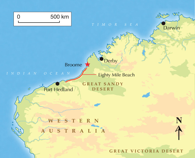
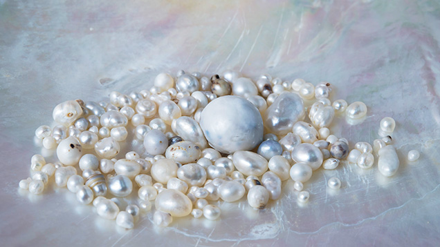
In late September 2013, a GIA pearl team (authors AH and AM) with the assistance of the Paspaley Pearling Company, once again had the opportunity to visit the historical P. maxima mollusk beds off Eighty Mile Beach in Broome, Western Australia (Scarratt et al., 2012, figure 1). Although the main purpose of the pearling expedition off Eighty Mile Beach was to collect wild P. maxima shells from the ocean floor as a part of the Australian shelling quota system (WAMSC, 2015), GIA’s primary objective was to collect natural pearls found in the mollusks. In total, 774 natural pearls were found in 20,488 opened mollusks (figure 2), and those authors present were able to retrieve pearls and record the exact locations where 370 of them formed within the mollusk (B1 sample type).
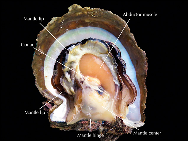
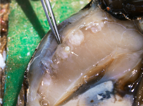
On opening the shells, the search started with a visual inspection of the soft organs for any obvious pearls. This was followed by a fingertip search to feel for any seed pearls or pearls that might have formed in areas hidden from view by the mantle that extended around the lip, down to the hinge and the central part (figure 3). The adductor muscle was also checked for the presence of pearls. The majority of pearls retrieved from the known positions were found in various parts of the mantle, either close to the lip, toward the hinge or in the central part of the mollusk (table 2). However, it was very interesting to see that some seed pearls were also found embedded in the adductor muscles (figure 4). As would be expected for natural wild shells, no pearls were found in the gonads. Moreover, a few were loosely attached to the shell surface, and were subsequently removed by applying gentle finger pressure. The latter are considered to be whole pearls and typically show circular marks of organic-rich material on their surface where they were connected to the shell (Lawanwong et al., 2019). These findings are in keeping with the observations from Australian fisheries recorded by Kunz and Stevenson (1908):

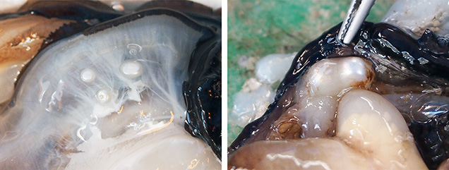
It came as no surprise that some specimens did not produce any pearls, while others contained more than one pearl. More commonly, pearls formed in individual sacs in adjacent areas (figure 5, left) or in separate areas within the mollusk, though in some cases several individual pearls formed within the same sac (figure 5, right).
Shell opening was carried out over eight days by several people on the vessel. The exact position in which some of the remaining 404 pearls formed inside the mollusks could not be documented because they fell out of the sac during the shell opening process (B2 sample type). This could have resulted from the abrupt retraction of the mantle lip from the shell edge (ventral margin) when cutting through the mollusk or from the perforation of the pearl sac by a knife. The balance of the 404 pearls were recovered later in the process from the bottom of a container after the mollusks’ soft tissues had passed through a mechanical separator (B3 sample type). Nonetheless, all the samples studied are undoubtedly classified as natural pearls of known origin, as they formed in wild mollusks (i.e., unbred, non-hatchery raised, unoperated) that had never been involved in any pearl culturing process. The measurements ranged from 0.49 mm to 16.16 × 15.57 × 13.24 mm, and the weights ranged from negligible to 19.94 ct.
The internal structures of the 774 natural marine pearl samples, on loan to GIA, were recorded using RTX and μ-CT analyses. The RTX analysis was performed using a Pacific X-ray Imaging (PXI) GenX-90P X-ray system with 4-micron microfocus, 90 kV voltage, and 0.16 mA current X-ray source with an exposure time of 200–400 milliseconds per frame, combined with a PerkinElmer 1512 flat panel detector with a maximum of 128 frames average and 74.8 micropixel pitch with 1944 × 1536 pixel resolution. The samples that showed intriguing or indistinct structures were selected for more detailed μ-CT work to better view the internal structures. The μ-CT work was carried out with a ProCon CT-mini X-ray system with a 5-micron microfocus, 90 kV voltage, and 0.18 mA X-ray current source. Two detectors with a frame grabber card were used to capture the results: a Hamamatsu flat panel detector C7921CA-29 with 50 micropixel pitch and 1032 × 1032 pixel resolution, and a Varex 1207 flat panel detector with 74.8 micropixel pitch and 1536 × 864 pixel resolution. RTX and μ-CT data were collected in GIA’s Bangkok laboratory. Because the work focused on the samples’ internal structures, other aspects including physical, spectroscopic, and chemical characteristics are not presented.
OBSERVATIONS AND RESULTS
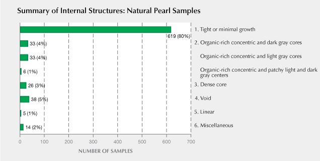
A natural pearl is the result of an accidental occurrence during the normal life cycle of a mollusk (Strack, 2006). Various conditions influence the formation of pearls, such as water environment and a mollusk’s health (Gervis and Sims, 1992; Bondad-Reantaso et al., 2007), so it is common to find various types of internal structures in natural pearls, as well as cultured pearls. Based on the internal features obtained by RTX and μ-CT analyses, natural pearl samples can be separated into six broad growth structure types (figure 6):
- Tight or minimal growth
- Organic-rich concentric
- Dense core
- Void
- Linear
- Miscellaneous
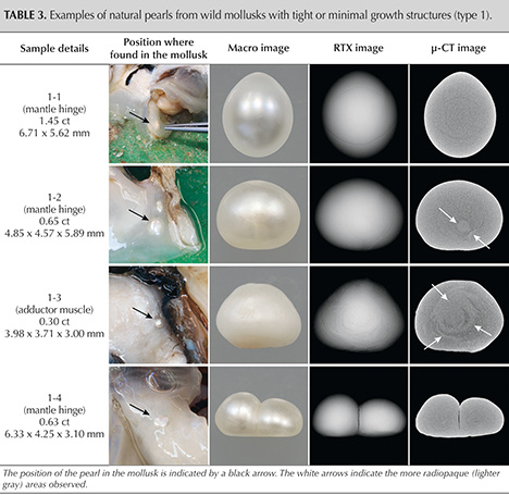
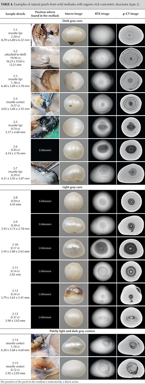
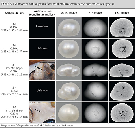
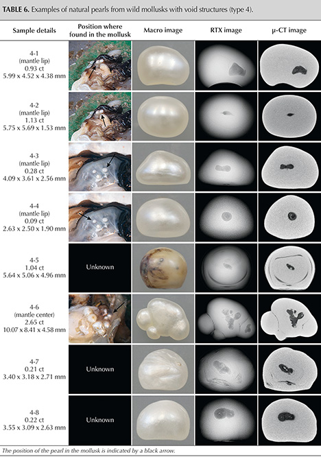
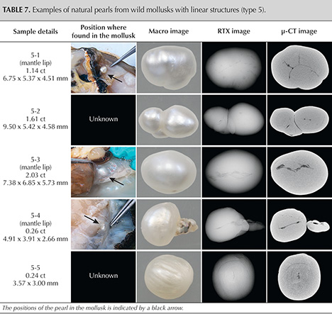
From all 774 natural pearls examined in this study, 45 were selected in order to provide a representative overview of the various internal structures observed. The RTX and μ-CT results of each sample are shown in tables 3 through 8, along with the sample number, weight, and measurement in the first column and a macro image in the third column. The second column shows the image captured during each pearl’s retrieval, if applicable, from within the mollusk. The position of each pearl is indicated by a black arrow and noted in the first column. Those listed as “unknown” were the ones where the pearl’s position could not be documented because it fell out of the pearl sac during the opening process or was later recovered from the mechanical separator. The RTX and μ-CT results are grayscale images in which differing shades of bright and dark grayscale intensity correspond to X-ray density (i.e., the different degrees of attenuation the component materials have to X-rays). Mineralized materials such as aragonite or calcite are denser or more radiopaque than organic-rich features or voids filled with gases and/or liquid; hence, an area composed entirely of aragonite will appear lighter than an area containing organic-rich or void features which generally appear darker (Wehrmeister et al., 2008; Sturman, 2009; Otter et al., 2014). [Note: In this article, the description of an internal structure as “dark gray” and “light gray” corresponds to its X-ray density, not its actual color.]
Type 1: Tight or Minimal Growth Structures. 619 samples (~80%) recovered displayed this structure. They lacked any clear internal growth structures, or only showed a few weak growth patterns (fine structural curved lines correlating with the pearl’s shape) by RTX analysis (table 3). The majority of the pearls also revealed very little structure when examined by μ-CT analysis. In addition, slightly lighter gray (more radiopaque) areas were observed in some instances, as shown in samples 1-2 and 1-3 (indicated by white arrows). These areas may be composed of some material or substance that has a higher X-ray density than nacre. Since the pearls did not contain questionable internal features related to any type of cultured pearl formation, they would, in almost all cases, be identified as natural pearls.
Type 2: Organic-Rich Concentric Structures. Around 9% of the samples in this study displayed organic-rich concentric structures of various sizes and patterns (table 4). They ranged from examples with a very small core (sample 2-1) to those with large concentric layered areas occupying almost half of the pearl’s interior (sample 2-10). μ-CT analysis clearly revealed cores with different forms, such as a dark gray core (more radiolucent), a light gray core (more radiopaque), or varying degrees of contrast with patchy light and dark gray centers within the concentric structures. The dark gray cores are not empty spaces, but rather they are filled with a solid/semi-solid organic-rich phase (samples 2-1 to 2-7). The sizes of the dark gray cores varied from small spots to large irregular areas shown in samples 2-6 and 2-7. The light gray cores appeared to be dense material and had a radiopacity similar to that of the outer nacre; hence, they are probably CaCO3. These cores varied in size, with the majority of them appearing small and rounded, as shown in samples 2-8 to 2-12. Sometimes the shape and structures varied, as in sample 2-13, which showed an ovoid core and a linear-appearing feature running across its center (magnified in figure 7). The structure of sample 2-13 was inconsistent with the majority of pearls in the group; it has never been observed in known or “reportedly” natural samples in our experience and has not been noted in the literature. This unusual structure may be challenging to identify, especially in laboratory conditions when information on the pearl’s origin is unknown, and therefore greater care is needed in its interpretation. It could easily be misidentified as a NBC pearl since the majority of light gray cores observed in natural pearls are typically rounded, and off-round cores have been observed in known NBC pearls produced by the P. maxima mollusk (Manustrong et al., 2019). Moreover, the central feature within the core has a linear form, and such features have been observed in NBC pearls produced by various mollusk species. Similar linear features, although not exactly the same, are often considered sufficient evidence to classify pearls as NBC (Hänni, 2006; Sturman, 2009; Krzemnicki et al., 2010; Sturman et al., 2016; Nilpetploy et al., 2018a; Manustrong, 2018). RTX and μ-CT imaging of samples 2-14 and 2-15 showed patchy concentric structures that appeared neither light nor dark gray, and these were due in part to differences in the organic content within the areas. These samples also lacked dark or light gray cores.

Type 3: Dense Core Structures. A small portion, approximately 3% of the group, contained solid light gray (more radiopaque) core features, usually single but sometimes multiple, at their centers (table 5). The X-ray attenuation of these cores closely matched some similar features observed in type 2 samples. However, they were not enclosed by clear organic-rich concentric structures; hence, they were classified as a separate structural type, referred to here as a “dense core.” These dense cores usually appeared as a single structure that was round, off-round, or irregularly shaped, and they were often associated with some faint organic-rich layers (samples 3-1 to 3-3). A few examples, such as samples 3-4 and 3-5, showed two or more cores that formed together as multi-nuclei. Irregular dense core structures have only been observed in natural pearls, in the authors’ experience, and have not been reported in any NBC pearls. However, an off-round feature associated with the main core in sample 3-5 looks suspicious and could be construed as being a light gray CaCO3 “seed” feature sometimes found in NBC pearls (Krzemnicki et al., 2010, 2011; Nilpetploy et al., 2018a; Manustrong et al., 2019). Thus, if this pearl was tested without any supporting provenance, it could be interpreted as being NBC. Nevertheless, the greater detail revealed by μ-CT analysis showed faint growth layers inside the feature, in contrast to the CaCO3 “seed” features, which are usually tight and contain no structure (magnified in figure 8). In this example, where the internal structural characteristics overlap and do not correspond with the majority of structures observed in natural and NBC pearls, an inconclusive opinion may well result.

Type 4: Void Structures. When discussing the internal characteristics of pearls, voids usually refer to radiolucent features that are less dense or more transparent to X-rays, such as cavities or areas filled with gas and/or liquid phases. Voids generally appear as various shades of dark gray, and thus they may resemble organic-rich features in low-resolution X-ray images. Voids are commonly observed in saltwater NBC pearls, especially pearls from Pinctada species (Wehrmeister et al., 2008; Sturman, 2009; Krzemnicki et al., 2010; Otter et al., 2014; Sturman et al., 2016; Nilpetploy et al., 2018a; Manustrong, 2018; Al-Alawi et al., 2020). In many cases, voids are a key identification feature by which to differentiate NBC from natural pearls. Only ~5% of the group showed void-related characteristics, and most of these exhibited centrally positioned irregular void-like forms after RTX analysis (table 6). However, μ-CT analysis revealed that many of these voids were filled with fine organic-rich growth features (samples 4-1 and 4-3 to 4-6), and some showed small light gray features in the center of organic-rich structures (samples 4-7 and 4-8) rather than the apparently empty spaces usually observed in NBC pearls. The voids are also small relative to the size of the pearl, while those found in saltwater NBC pearls tend to be larger and more elongated, extended lengthwise, usually taking up more than half of the pearl’s length.
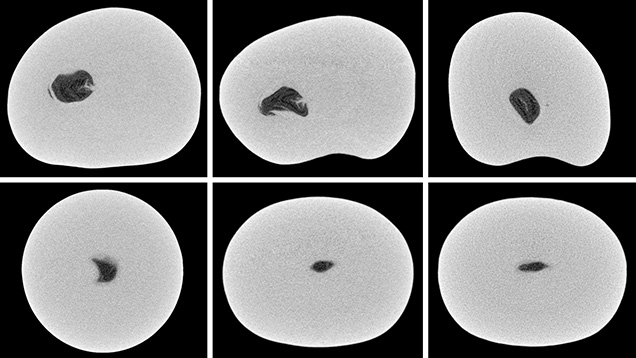
While natural voids do show some differences from those encountered in NBC pearls, identification challenges could cause some pearls to be misidentified. A couple of samples in this study showed voids similar to those characteristically found in NBC pearls, and they would almost certainly be classified as NBC if tested under blind conditions. It is interesting to note that samples 4-1 and 4-2 were retrieved from the same pearl sac and both possessed void features. However, the void in sample 4-2 does not appear to be filled with organic-rich growth structure and contains fewer light gray linear features, a common observation in NBC pearls (magnified in figure 9).
Type 5: Linear Structures. Most linear structures are thin voids, with or without organic-rich material partially filling them. Owing to the different appearances of both types of formation, they are separated into different groups in this article. Linear structures have been regarded as characteristic of NBC pearls from both saltwater and freshwater environments (Scarratt et al., 2000; Sturman, 2009; Krzemnicki et al., 2010; Sturman et al., 2016; Nilpetploy et al., 2018a; Manustrong, 2018; Al-Alawi et al., 2020).
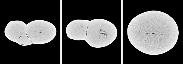
Interestingly, 5 of the 774 natural pearl samples also displayed linear structures (table 7). Sample 5-1 showed a complex linear feature corresponding, to some degree, with the pearl’s baroque outline. In the authors’ experience, such linear structures may be characteristic of NBC pearls, but this example demonstrates that is not always the case. Sample 5-2 consisted of three portions, two of them having a long linear feature at the center (magnified in figure 10). Although the other portion only displayed a few growth patterns, the pearl’s overall structure looked questionable and it could easily be misidentified as an NBC pearl. The linear structures revealed in samples 5-3 to 5-5 are quite distinct and correspond with the length of the pearl, a characteristic shown by many NBC pearls.
Type 6: Miscellaneous Structures. The final group of natural pearls were classified as those with “miscellaneous structures.” This group includes samples possibly containing marine organisms entombed by nacre deposition, examples containing obvious internal fractures, and pearls exhibiting a mixture of structures that do not readily fit into any of the former five structure types listed (table 8).
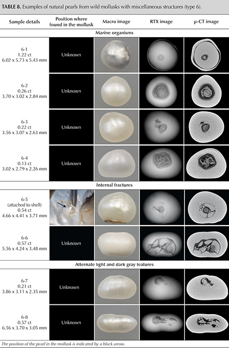
Pearls with internal features that appear to be related to marine organisms are very rare, and only a few have been encountered in the GIA laboratory from the thousands of natural pearls submitted for identification and research. Some examples have been documented in the existing literature (Scarratt et al., 2012; Nilpetploy [nee Somsa-ard], 2015). Even so, the authors were surprised to find four samples in this study group with what are most likely marine organism features. Samples 6-1 to 6-3 revealed shell-like features at their center that likely initiated the formation of the pearls, together with displaced epithelial cells. The features measured 0.78 × 0.70 mm, 1.46 × 1.34 mm, and 0.72 × 0.50 mm in diameter, respectively. The resulting pearls are similarly small. The exact identity of the marine organisms is unknown; based on the morphology observed by μ-CT analysis, however, they could be one of the many benthic foraminifera species. Foraminifera are single-celled organisms (protists) with a test (shell) that can possess one or more chambers (Borrelli et al., 2018). They may be composed of organic compounds, sand grains, or other particles cemented together, or crystalline CaCO3, and are usually less than 1 mm in size (Steele et al., 2001). One of the existing references (Nilpetploy [nee Somsa-ard], 2015) described what was thought to be an intricate foraminifera sphere observed in a 0.95 ct natural pearl. The core feature was encircled by a dark organic-rich area and accompanying void similar to the natural pearls described here. Sample 6-1 (magnified in figure 11) has a distinct outer ring that looks questionable and could be considered diagnostic of an atypical bead cultured pearl (aBCP), but it does not contain an “organic tail” typically observed in aBCP pearls (Hänni et al., 2010; Scarratt et al., 2017; Kessrapong and Lawanwong, 2020). Therefore, it would be identified as a natural pearl, especially when its size and quality are also taken into account. Sample 6-4 displayed a large irregular radiopaque material together with some small fragments at its center. As the magnified images in figure 12 show, the material’s structure differs from the three former samples, though its appearance is also likely that of a marine organism. The main feature, with its porous-looking pattern, bears some similarity to the structure of Melithaea, a genus of colonial soft coral (also known as fan coral), which is distributed throughout the tropical Indo-Pacific region (Jeong et al., 2018; Atlas of Living Australia, 2020).


Samples 6-5 and 6-6 displayed severe internal fracturing at the center of the features. X-ray computed tomography clearly revealed the intriguing structures of both pearls and indicated they were “fragmented.” It is puzzling how such broken features can exist within known natural pearls whose overlying layers of nacre appear intact and where no drilling or external treatment of any kind exist. Sample 6-6 is especially perplexing, as the naturally formed concentric organic-rich inner structure has disintegrated into many parts (magnified in figure 13) to fill the void present. This intricate structure is highly unusual, and it would be challenging to correctly identify such a pearl in laboratory testing conditions. Some of these samples could be suspected of being the result of culturing using various atypical nuclei. Yet their generally very small size would suggest otherwise, and it is known that this is definitely not the case, as they are confirmed natural pearls.

Samples 6-7 and 6-8 revealed a mixture of structures alternating between more radiopaque (light gray) and more radiolucent (dark gray) features. The structure exhibited in sample 6-7 (magnified in figure 14) appears to take the form of two initiation features merging together. Although the structure is unusual, this sample would almost certainly be identified as a natural pearl since there are no characteristic cultured pearl features such as a void or a linear structure in the center. On the other hand, sample 6-8 (magnified in figure 15) displayed a complex elongated structure consisting of a linear-like feature at its center combined with dark gray areas of voids and organic matter, as well as other solid light gray areas. Such a structure looks suspicious, and in laboratory conditions the pearl could be misidentified as NBC.


DISCUSSION
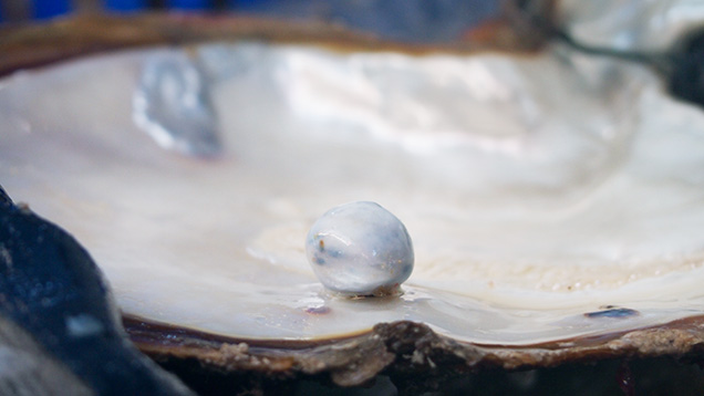
Pearl identification mainly involves separating pearls that formed naturally (figure 16) from those that resulted from a culturing process. RTX is the primary technique used in gemological laboratories to examine a pearl’s internal structure, while μ-CT is a high-resolution 3D imaging technique that is sometimes used to visualize internal structures in greater detail. Interpreting the structural results obtained depends on the research and data collection of known reference samples by well-trained, experienced gemologists. Studying this large group of natural pearls provided a greater understanding about the range of microradiographic structures encountered in natural pearls produced by wild P. maxima mollusks. The results showed that most of the natural pearl samples (~80%) exhibited tight or minimal growth structures (type 1). These pearls are straightforward to identify as natural. This observation is also consistent with the majority of natural pearls tested in all GIA’s pearl testing locations. Tight or minimal growth structure is commonly observed in natural pearls produced by many mollusk species of both nacreous and non-nacreous pearls (Krzemnicki et al., 2010; Scarratt et al., 2012; Karampelas, 2017; Nilpetploy et al., 2018b; Scarratt and Hänni, 2004; Wing Yan Ho and Zhou, 2014; Wing Yan Ho, 2015; Wing Yan Ho and Yazawa, 2017).
Organic-rich concentric structures (type 2) are also known to be typical of natural pearls produced by various mollusk species (Krzemnicki et al., 2010, Scarratt et al., 2012; Sturman et al., 2014; Karampelas et al., 2017; Nilpetploy et al., 2018b), yet they were found in only about ~9% of the samples. This includes organic-rich concentric and dark gray (radiolucent) cores that are filled with organic-rich features (~4%), organic-rich concentric and light gray (more radiopaque) cores in which the majority of the cores are small and rounded (~4%), and organic-rich concentric and patchy light and dark gray structures lacking dark and light gray cores (~1%) (see figure 6). Most of the organic-rich concentric areas observed in natural pearls cover a relatively small portion of the pearl’s interior. It is important to note that none of the pearls in this study showed any of the light gray CaCO3 “seed” features reported by Manustrong et al. (2019; see tables 4 and 5) and previously reported by other authors (Krzemnicki et al., 2010, 2011; Nilpetploy et al., 2018a) as being associated with organic-rich concentric structures in some cultured pearls. At the time of this report, the irregular dense core structures (type 3) free of any clear organic-rich concentric features are only found in natural pearls and have not been reported in NBC pearls in the literature.
Voids (type 4) and linear structures (type 5) have been reported as characteristic features in P. maxima NBC pearls (Sturman et al., 2016), but they were found in only ~5% and ~1%, respectively, of the natural pearl samples in this study. Considering that the internal structural features of natural and NBC pearls may overlap, the separation between the two can be challenging and their interpretation requires extra care. While the pearls’ external appearance may also assist in their identification, it is a subjective element and can vary greatly depending on the gemologist’s experience. Therefore, the internal structures of pearls provide the most important evidence and consequently are the primary means of identification. In general, the void structures observed in the natural pearl samples studied occupy a relatively small volume of the pearl, and are often filled with organic-rich growth structures. The linear features are perhaps the most questionable structures found in natural pearls since the structure is widely accepted as being an identifying feature of NBC pearls produced by various mollusks. Therefore, it is possible that some or all of the five natural pearls that revealed linear structures would be identified as NBC if submitted on a “blind” basis.
In the miscellaneous structures (type 6), the presence of a marine organism-like feature as a nucleus in four samples emphasized a frequently applied hypothesis that a natural pearl forms when an external foreign object reaches the mollusk’s mantle (epithelium layer), and causes the epithelial cells to encroach into the connective tissue and enclose the object to create a pearl sac, which secretes the necessary components (Kunz and Stevenson, 1908; Landman et al., 2001; Strack, 2006; Southgate and Lucas, 2008). However, this is a rare occurrence, as only a very small number of pearls have reportedly shown this type of structure, and there are several theories about how natural pearls may form. These include: forming as a result of injury to, or abnormal growth of the epithelium cells; epithelial cells separating to form pearl sacs of their own accord; and health-related instigation. A concept mentioned in Strack (2006) may help to explain the severe internal fracturing of the features within the centers of samples 6-5 and 6-6:
Lastly, the relationship between the internal structure and the position in which a pearl forms inside the P. maxima mollusk species is not entirely clear at this time. Since it was not possible to record the position in which over 50% of the samples were found in the mollusk’s body, this question could not be addressed. Such a study would require a greater sample set of pearls with suitably accurate records. However, it was noted that most of the pearls found in the adductor muscles possessed tight or minimal growth structures, while mantle pearls generally showed a variety of structure types.
CONCLUSIONS
The results of this study show that various forms of internal structures are observed in natural pearls. The majority of samples revealed limited growth features (minor growth patterns) or no clear structure at all (tight structure), confirming their natural identity. The relatively few samples that exhibited varying degrees of structural overlap, internal characteristics possibly observed in NBC pearls or natural pearls (i.e., off-round light gray cores in concentric organic-rich structure, voids, and linear features), raised concerns about the identification of such pearls when tested without any supporting provenance information. They demonstrated the complexities often faced with pearl identification. It is not unheard of for the same pearl to receive different determinations from different organizations based on the equipment and techniques used to obtain the data, the experience and specialized knowledge of gemologists interpreting the data, and the comprehensiveness of the reference sample database. Natural pearls containing either a large void relative to the pearl’s size or a distinct linear feature have a greater probability of being defined as NBC. In cases where the structures are extremely complex or ambiguous, or do not conform with anything in the database, an “inconclusive” call is likely. GIA recognizes the ongoing pearl identification challenges and intends to continue studying the internal structures of known natural and cultured pearl samples from various environments and mollusks to strengthen its reference collection and database, thus ensuring GIA’s gemologists have the most up-to-date information at hand before reaching any conclusions on a client’s pearl submissions.


