Fluorescence Spectroscopy for Colored Pearl Treatment Screening
Color is one of the most important value factors for pearls. Commonly applied treatment methods used to alter their color to increase commercial value include dyeing, irradiation, and bleaching. Unfortunately, the identification of some color treatments is challenging and time consuming. In this study, we tested the use of a fluorescence spectroscopy system to nondestructively inspect pearls by measuring their near ultraviolet (UV) response to visible fluorescence under mid-UV excitation.
When excited by deep to mid-UV (200 to 300 nm) light, naturally colored pearls emit a fluorescence band between 320 and 400 nm centered at 340 nm. This fluorescence feature may be attributable to the organic compounds contained within the nacreous layers (J. Hiramatsu et al., “Non-destructive assessment of the effects of heat and sunlight on akoya pearl quality,” Seibutsu Kogaku, Vol. 88, No. 8, 2010, pp. 378–383; F.W.J. Teale, “The ultraviolet fluorescence of proteins in neutral solution,” Journal of Biochemistry, Vol. 76, No. 2, 1960, pp. 381–388). It can be identified in all untreated pearls, corresponding to the UV absorption band around 280 nm (J. Yan et al., “Origin of the common UV absorption feature in cultured pearls and shells,” Journal of Materials Science, Vol. 52, No. 14, 2017, pp. 8362–8369).
Commonly applied color treatments such as dyeing and irradiation tend to damage or mask the conchiolin in the nacre, significantly reducing the fluorescence intensity. By evaluating the intensity of a pearl’s fluorescence in the UV region, it is possible to rapidly detect potential color treatments on pearls in a nondestructive manner.
A prototype fluorescence spectroscopy system was designed to measure the fluorescence signal in order to detect potential color treatments. A 275 nm UV light-emitting diode (LED) was used as the excitation source. The excitation light was guided by a bi-fabricated fiber probe to generate the fluorescence signal from the pearl sample, and the fluorescence signal was relayed by the same fiber probe to the detector. Finally, a spectrometer was used to disperse the fluorescence light emitted and monitor the response in the 300 to 700 nm range.
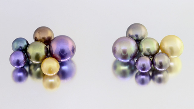
A set of 12 pearl samples was selected for evaluation. Figure 1 shows six treated-color pearls (left) and six naturally colored pearls (right). The samples included both freshwater and saltwater cultured pearls of colors frequently encountered in the market.

Figure 2 shows the experimental results of this fluorescence measurement prototype. The horizontal axis indicates the fluorescence wavelengths from 300 to 550 nm while the vertical axis shows the normalized detector counts, which is the relative intensity of the signal normalized to the spectrometer’s integration time per millisecond. Based on the results, the six naturally colored pearls showed fluorescence signals in the UV region at least 2.5 times stronger than the six treated pearls. This characteristic feature may be a useful and rapid screening technique for gemological laboratories to detect color treatments in pearls.


