Pleochroism and Color Change in Faceted Alexandrite: Influence of Cut and Sample Orientation
ABSTRACT
The color appearance of faceted gemstones is a complex subject, and the challenges are increased if the material is biaxial and pleochroism is added to the considerations. For alexandrite in particular, the quest for a beautiful cut gem is further intensified by efforts to achieve the “best” color change. As an optically biaxial material, alexandrite possesses three different vibration directions X, Y, and Z. These are parallel to the three crystallographic axes a, b, and c, each of which has a distinct pleochroic color.
The present study seeks to evaluate the effect of various factors on color and color change using two groups of faceted synthetic alexandrites of comparable sizes and cuts with table facets oriented perpendicular to one of the three crystallographic axes. If the faceted gemstones are examined in transmitted light in immersion with a polarizer between the sample and the observer, the basic pleochroic colors can be separated and seen individually. For the synthetic alexandrites, if the faceted gemstones are examined in reflected light, this study demonstrates that the mixing of the three colors X + Y + Z, caused by multiple reflections of light within the faceted stones, greatly diminishes the role of table facet orientation on the quality of color and color change in well-cut gems. Likewise, for other biaxial stones it is expected that the effects of pleochroism will also be reduced in faceted stones to some extent.
INTRODUCTION
Alexandrite, the chromium-bearing variety of chrysoberyl, shows distinct pleochroism and a signature color change between daylight (or daylight-equivalent fluorescent light) and incandescent light, referred to as the alexandrite effect. A similar color change is also observed for other varieties of gem minerals such as garnet, sapphire, spinel, kyanite, fluorite, and diaspore (Bosshart et al., 1982; Gübelin and Schmetzer, 1982; Schmetzer et al., 2009).
Chrysoberyl, belonging to the orthorhombic crystal system, is birefringent and optically biaxial. If unpolarized light enters a birefringent crystal, the beam is split into two polarized waves in all directions not parallel to an optic axis. These two waves leave the crystal in polarized form and can be separated, and seen individually, by rotating a polarizer (i.e., a polarizing filter) located between the sample and the observer. The optically biaxial nature of chrysoberyl further means that the optical indicatrix has three different vibration directions X, Y, and Z, which are parallel to the three crystallographic axes a, b, and c (Bloss, 1961; Wahlstrom, 1969; Kerr, 1977). In the three vibration directions, light can be differentially absorbed, and an absorption spectrum can be measured for each direction. These three directions thus generate three basic pleochroic colors (Burns, 1993; Schmetzer and Bosshart, 2010; Schmetzer et al., 2012, 2013; Sun et al., 2017; see also Devouard and Notari, 2009).
In views parallel to one of the three a, b, or c1 crystallographic axes in alexandrite, two vibration directions and two of the three basic colors are always present simultaneously, i.e., X + Y, X + Z, or Y + Z (figure 1). Stated otherwise, the color seen with the unaided eye when looking parallel to any of the three crystallographic axes is always a mixture of two of the X, Y, and Z basic color components (Schmetzer and Bosshart, 2010; Schmetzer and Malsy, 2011).
The specific colors observed (and the spectra produced) are in turn dependent in large part on the concentrations of color-causing trace elements and the path length of light through the crystal. Principal trace elements affecting color in alexandrite are chromium, vanadium, and iron. Variations in path length result in the optical phenomenon referred to in gemology as the Usambara effect, with color changing or shifting as the path length increases (Halvorsen and Jensen, 1997; Halvorsen, 2006).
The color impressions just described, based on visual appearance and examination, have also been evaluated and verified through colorimetric measurement of natural alexandrite crystals, natural alexandrite cubes, and synthetic alexandrite cubes with different trace-element contents (Schmetzer and Bosshart, 2010; Schmetzer and Malsy, 2011; Schmetzer et al., 2012, 2013). For instance, colorimetric measurements using oriented cubes of synthetic alexandrite with edge lengths from 2 to 10 mm (Schmetzer et al., 2013) demonstrated that, regardless of size, for all three different orientations parallel to the a-, b-, and c-axes, changing between daylight and incandescent light resulted in a respective increase or decrease in blueness and redness (the alexandrite effect). With increasing cube size, a color shift was visible. The larger the cube, the redder the alexandrite appeared in both daylight and incandescent light (the Usambara effect).
The foregoing optical characteristics and phenomena have long spawned efforts to ascertain preferred orientations for fashioned alexandrites (and pleochroic materials more broadly). For alexandrite in particular, the goal of gem cutters and merchants for decades has been the “best” color change, defined as a green or bluish green to blue-green color in daylight and a red-purple or reddish purple to purple color in incandescent light. Conventional wisdom among cutters has traditionally held that this favored color change is obtained if the table facet is oriented perpendicular to the b-axis (Fischer, 1954). In practical terms, however, cutting in such a direction could prove problematic at times on account of the cyclic twinning commonly seen in natural alexandrites.
The above-noted work with oriented crystals and cubes has also lent scientific support to the traditional understanding, concluding on the basis of visual inspection in transmitted light and colorimetric measurements that the “best” and most highly desired color change between daylight and incandescent light was observed in a direction of view parallel to the b-axis (Schmetzer and Bosshart, 2010; Schmetzer and Malsy, 2011; Schmetzer et al., 2012).
Hughes (2014) described pleochroism using a simplified theoretical model for light behavior in optically biaxial faceted gemstones. That model was based upon a single light beam entering the crystal perpendicular to the table facet. If the table facet were oriented perpendicular to one of the crystallographic axes, that beam would be split into two of the three basic components X, Y, and Z. If next reflected from pavilion facets, the light beam would travel through the crystal in a direction parallel to another of the three crystallographic axes and, consequently, would contain the third basic color component. After another reflection at the pavilion, the light leaving the faceted gemstone would be a mixture of all three components X, Y, and Z.
Hughes (2014) further noted that, depending on the cut of the sample, the light path length could vary based on whether the beam entered the faceted gemstone near the center of the table facet or near the girdle. Hence, the mixture of light reflected from different pavilion facets would show different percentages of X, Y, and Z, thereby generating different colors. Unfortunately, however, no faceted biaxial gemstones with known orientations were presented to support the theoretical model.
More recently, Sun et al. (2017) both inspected visually and measured colorimetrically a solely Cr-bearing Czochralski-grown synthetic alexandrite cuboid with edges between 2.65 and 3.18 mm and also calculated colorimetric data maps detailing color, chroma, chroma difference, hue angle difference, and color difference for wafers in various orientations and with path lengths between 1 and 25 mm. One point explicitly highlighted was that areas with large values for hue angle difference or color difference did not necessarily show the “best” orientation for the desired color change.
Maps of colorimetric data were likewise calculated for faceted alexandrites. Based upon the general considerations of Hughes (2014) for optically biaxial gemstones, Sun et al. computed parameters for color and color change for a hypothetical faceted stone with a 10 mm light path length. Using preferred ranges for hue angle for daylight versus incandescent light and large chroma values for both light sources for alexandrite, these authors tried to find the “best” orientation of the table facet or, in their own words, to “orient a stone along the ‘best’ direction.” It was concluded “that pleochroism in a faceted gemstone serves to smear out the “best” direction for color change.” Furthermore, it was found “that stones cut with their table to culet direction oriented perpendicular to the b-axis show the best color change, while orientation parallel to the b-axis produces weaker color change” (Sun et al., 2017; Z. Sun pers. comm., 2018).2
Again, however, no faceted stones were examined and compared with the results obtained by theoretical calculations of colorimetric parameters.
Thus, with regard to faceted alexandrites found in the trade, the current situation remains one where questions abound. Due to differences in trace-element content, sample orientation, size, and cut, numerous parameters exist that might influence color and color change. The present study therefore attempts to address queries involving the influence of these factors using carefully prepared samples of faceted material.
SAMPLES
The high value of facet-quality natural alexandrite material renders it nearly impossible to obtain suitable rough for cutting several small samples with different known orientations from the same large rough crystal. Hence, the present study was performed with synthetic gem material. Two groups of three samples each were cut from two synthetic crystals. An overview is provided in table 1.

One group consisted of three samples cut from a flux-grown synthetic alexandrite produced by Creative Crystals Inc. in San Ramon, California (see Schmetzer et al., 2012). The crystal was grown with a seed oriented parallel to b (010), and square or almost square table facets were cut parallel to either a (100), b (010), or c (001). Simple step cuts (emerald cuts) were fashioned with a table facet, three rows of crown facets, and three rows of pavilion facets (figure 2). Sizes ranged from 8.3 × 8.1 × 7.0 mm to 9.1 × 8.6 × 8.2 mm.

The other group comprised three synthetic alexandrites faceted from a crystal grown by the HOC technique in Novosibirsk, Russia, by V.V. Gurov (see Schmetzer et al., 2013). Starting with pieces sawn from the rough crystal, likewise produced with a seed parallel to a (010), table facets were again oriented parallel to either a (100), b (010), or c (001). An oval brilliant cut was used for the crown, with a mixed cut of brilliant and step facets for the pavilion (figure 2). Sizes spanned from 8.0 × 6.0 × 4.2 mm to 8.1 × 6.1 × 4.2 mm.
Within each group, the identical cuts and similar dimensions enabled a direct comparison of the influence of cut orientation upon color and color change.
CHEMICAL PROPERTIES
The flux method employed by Creative Crystals for the alexandrites examined here used a series of several sequential growth cycles. As a result, the amount of chromium and iron, the principal color-causing trace elements in these samples, incorporated in each growth layer was variable. Mean values ranged from 0.18 to 0.26 wt.% Cr2O3 and from 0.86 to 1.14 wt.% Fe2O3. Vanadium contents were approximately 0.01 wt.% V2O3 (Schmetzer et al., 2012).
The synthetic alexandrites grown by the HOC technique, in contrast, were more homogeneous in chemical composition, with chromium measuring from 0.30 to 0.43 wt.% Cr2O3 and vanadium ranging from 0.07 to 0.14 wt.% V2O3. Iron levels were at 0.01 wt.% Fe2O3 or below (Schmetzer et al., 2013).
VERIFICATION OF SAMPLE ORIENTATION
The orientation of the table facets, which were cut according to morphological features of the rough gem alexandrites, was verified by ascertaining the positions of the optic axes in each stone. The optic axes lie in the optic plane, which in chrysoberyl is the ac-plane, and the c-axis is located exactly between the two optic axes (figure 3). The b-axis is perpendicular to the optic plane. Consequently, by viewing in a direction parallel to the optic plane and rotating a sample about the b-axis, both optic axes may be brought parallel to the direction of view.

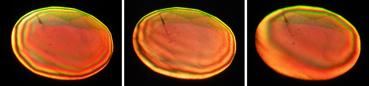
In applying this information to the flux material grown by Creative Crystals, the task was aided by growth planes visible in immersion parallel to the seed (010) and, in one sample, additional growth planes parallel to the prism k (021) (figure 4). By using that insight (in conjunction with the observed pleochroism; see below), it was possible with reasonable ease to find the b-axis of the crystals and to use that axis for rotation in the immersion microscope. In so doing, an interference pattern consisting of several rings would be obtained if an optic axis were slightly inclined to the direction of view. Tilting the faceted alexandrite toward a position in which the optic axis was parallel to the direction of view would then move the interference rings toward the center of the sample. The positions of both optic axes in the optic plane of the gemstone could thus be located, leading directly to the positions of the crystallographic axes a, b, and c and making apparent the orientation of the table facet relative to the crystallographic axes. The practical measurements were made by means of two- and three-axial sample holders with attached dials to measure angles. For all three step-cut samples, the deviation of the table facets from the intended orientation was below 5°.

The same procedure was applied for the three oval samples cut from a crystal grown by the HOC technique. Because these crystals, in general, did not show distinct growth planes, finding the proper orientation for the sample in the immersion microscope with the b-axis as the rotation axis was somewhat more time consuming. However, after locating both optic axes through observation of interference patterns (figure 5), the positions of the three crystallographic axes were determined, and the inclination of the table facet to the relevant crystal axis was measured. It was again found that the deviation of the table facets from the intended orientation was below 5° for all three mixed-cut gemstones.
COLOR BEHAVIOR OF FACETED GEMSTONES IN TRANSMITTED LIGHT
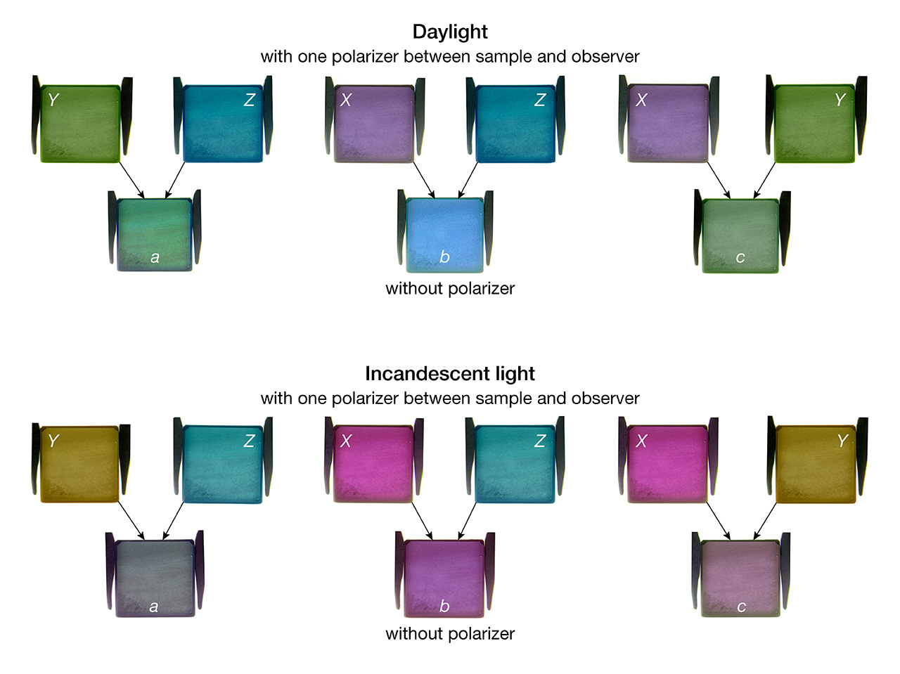
As noted at the outset, unpolarized white light in birefringent chrysoberyl crystals is split into two polarized waves, which can in turn be separated and seen individually by rotating a polarizer placed between the sample and the observer. For purposes of evaluating this phenomenon in faceted alexandrites in transmitted light, the samples were observed in immersion. By doing so, reflection of light at the pavilion facets and the corresponding mixing of different color components could be largely avoided. The only minor side effect of the methodology was a slight shift in color toward yellow on account of the immersion liquid.
All six oriented samples displayed the pleochroic behavior and colors commonly seen in alexandrites of similar size in both daylight and incandescent light, as follows (figure 6):
Daylight: X || a = violet-purple, Y || b = yellow-orange, Z || c = intense blue-green
Incandescent light: X || a = reddish purple, Y || b = orange, Z || c = green

The results were consistent with the established orientation of the table facets of the six samples as described above. The colors of X, Y, and Z observed visually were almost identical for the different samples of each group (flux-grown and HOC-grown synthetic alexandrites), with no distinct differences. Likewise demonstrated was the effect of differences in light path length and stone thickness, insofar as a fading of the color from the center of the table (or the culet) to the girdle was seen, especially in the step-cut samples grown by Creative Crystals.
Still in transmitted light but without a polarizer, two of the three basic colors previously separated by means of the filter were mixed in each direction of view. In all three directions, a color change between daylight and incandescent light was perceived (figure 7):
Daylight: view || a = green, view || b = blue-violet, view ||c = greenish yellow
Incandescent light: view || a = red-purple, view || b = reddish purple, view || c = red-purple

Again, no distinct differences were seen when comparing samples with the table facets in the same orientation, regardless of the cut.
Thus, to summarize, in transmitted light and especially in immersion, it was possible to observe different colors in views parallel to one of the three crystallographic axes and, by means of a polarizer, to separate the three basic colors of X, Y, and Z.
COLOR BEHAVIOR OF FACETED GEMSTONES IN REFLECTED LIGHT
Turning to the scenario in reflected light, the impression was one of a mixture of all three color components. To evaluate the general underpinnings of this situation, the simplified model presented by Hughes (2014) for biaxial gemstones in general was applied to the synthetic alexandrites examined.
With regard to the samples faceted with a relatively simple step cut (emerald cut), and by neglecting the refraction of light entering the crown facets and assuming a pavilion angle (the angle between the table and a pavilion facet) of 45°, a similarly simplified model for the path of light could be drawn. An example is given in figure 8 for an alexandrite cut with the table facet perpendicular to the b-axis. Unpolarized white light entering the sample at (1) through the table and crown facets would travel along the b-axis and split in the sample into X and Z color components. After reflection at the pavilion, light would travel along the c-axis, with X and Y color components. After a further reflection at the opposite side of the pavilion, the light beam would again travel along the b-axis. Light leaving the sample at (3) would thus contain X, Y, and Z color components. Reversing the scheme, light entering the sample at (3) would traverse along analogous paths, leaving the sample at (1). Light paths are not shown for this example.

If the starting point is shifted by 90°, light entering the sample at (2) through the table and crown facts would travel along the b-axis and split in the sample into X and Z color components. After reflection at the pavilion, the beam would travel along the a-axis, with Y and Z color components. Following a second reflection opposite, the beam would return to traveling along the b-axis. Upon exit at (4), the light would consist of two polarized waves with each wave showing X, Y, and Z color components. A reversed scenario for light entering at (4) would display a similar route, leaving the sample at (2). Again, light paths are not shown for this example.
Hence, application of the simplified model reveals that for each light path just described, the beam leaving the sample at the table or crown facets would have X, Y, and Z color components. If the simplifying assumptions are removed from the equation, light entering the alexandrite would be refracted at the crown facets, and the beam traveling through the sample would be reflected at facets with pavilion angles other than 45° (figure 9). For example, the three pavilion angles of the step-cut alexandrites examined here measured 62°, 54°, and 42°, respectively. The actual behavior in the faceted step-cut alexandrites would therefore entail a far more diverse collection of beams, path lengths, and oblique directions through the sample. Such oblique directions, in turn, would alter the absorptions and corresponding mixtures of X, Y, and Z color components present in each beam exiting from the faceted gemstone. As a result, the light leaving the sample would show a more complex mixture of X, Y, and Z components than calculated on the basis of the simplified model.

Analogous theoretical considerations would pertain for samples cut with the table facets in other directions (i.e., perpendicular to the a- and c-axes). Similarly pertinent, if the cut is more complicated, such as that of the three HOC alexandrites with an oval brilliant-cut crown and a mixed-cut pavilion, more reflections would be generated, with light passing through the samples in an even greater number of different directions.
The foregoing theories suggesting extensive color mixing were tested on a practical basis using the six faceted samples in both daylight and incandescent light. The alexandrites of the two groups were placed table-up on a grooved plastic stone tray, which made it possible to view all six simultaneously while the light of a single lamp was reflected from their table facets. As such, the color impression given by each of the samples was quite similar, notwithstanding the different cut orientations for the table facets. This similarity thus corroborated the mixing of all three colors in faceted stones due to multiple reflections.
More specifically, with respect to the three step-cut alexandrites, some minor differences in color could be discerned. In daylight, the sample cut with the table facet perpendicular to the b-axis was blue-green, while the other two were less blue, more green, or even slightly yellowish green (figure 10). In incandescent light, the sample with the table perpendicular to the b-axis was intense purple, while the two other two were less intensely colored, somewhat more reddish purple or red-purple.

When using the stone tray arrangement, the colors of the three samples with a more complex cut and a greater number of crown and pavilion facets were equivalent to each other in daylight and incandescent light. No visual color differences were observed. Moreover, if the alexandrites were placed on a glass plate, with several light sources illuminating them from different directions through the table and crown facets, or if the alexandrites were observed in diffused daylight, the differences in visual impression for even the step-cut samples became almost indistinguishable.
The divergence of the above results from the colors calculated by Sun et al. (2017) for alexandrites of various orientations are potentially explained by the simplifications employed for the model used there. In particular, the calculations neglected refraction of light at the crown facets, reflection of light from pavilion facets at angles of other than 45°, and especially multiple reflections from light entering the stone at directions oblique to the table facet. As a consequence, oblique directions of travel were not taken into account, yet these contribute significantly to the lively appearance of a faceted gemstone. It would be interesting to see if an augmented, more complex model incorporating a more realistic group of reflections and light paths within the sample would yield a closer approximation of reality.
In summary, the practical observations in the present study indicate that the cut and the orientation of the table facets play some role in achieving the desired colors in daylight or incandescent light. Nonetheless, all three simple step-cut alexandrites showed the “desired” color change. For samples with a more complex cut, such as a brilliant or mixed cut with numerous reflecting pavilion facets, the orientation of the table facet in such alexandrites is an almost negligible factor in determining color and color change.
CONCLUSIONS
Examination of two groups of faceted synthetic alexandrites with three known specific orientations of the table facets confirmed the general applicability of widely held tenets regarding color in the gemstone and also showed that as the cuts become more complex, such considerations have a diminishing effect on the actual appearance. In alexandrite, the three main color components of X, Y, and Z are quite different, but their visual impact can depend heavily on the viewing scenario.
When faceted samples are observed in transmitted light, with or without the use of a polarizer, distinct color differences dependent on the orientation of the table facets can be perceived. Conversely, in reflected light, the color differences become less discernible. More particularly, the differences are weaker in samples with a simple step cut and nearly nonexistent with a complex cut.
Thus, to summarize the progression in moving from basic cases to the more complicated (see also Schmetzer et al., 2013):
- Cubes of different sizes viewed in transmitted light parallel to the a- and c-axes are green or even slightly yellowish green in daylight and red-purple or reddish purple in incandescent light. Cubes viewed parallel to the b-axis are more blue-green, blue, or even violet in daylight and more purple or purplish violet in incandescent light. In other words, parallel to the a- and c- axes there is a stronger yellow component in daylight and a stronger red component in incandescent light, while parallel to the b-axis there is a stronger blue component in both daylight and incandescent light.
- Faceted alexandrites with a simple cut such as a step cut exhibit, when viewed in reflected light, behavior similar to that of the cubes, albeit potentially weaker. Those with table facets perpendicular to the a- and c-axes are green or even slightly yellowish green in daylight and red-purple or reddish purple in incandescent light. Those with table facets perpendicular to the b-axis are more blue-green, blue, or even violet in daylight and more purple or purplish violet in incandescent light. The differences diminish with multiple light sources at a variety of angles, or with diffused light.
- Faceted alexandrites with a complex cut—such as a modern brilliant or mixed cut—display, when viewed in reflected light, almost no discernible color difference dependent upon table facet orientation.
The faceted samples studied here also demonstrated the interrelated effect of trace-element concentration and light path length. To wit, although the flux-grown alexandrites were larger than those grown by the HOC technique, the intensity of their colors was similar because the sum of color-inducing trace elements in the HOC material—primarily chromium and vanadium—was approximately double that in the flux material. The longer light path lengths in the three larger flux-grown samples (between 7.0 and 8.2 mm from table to culet) broadly compensated for the higher concentration of chromium and vanadium in the smaller HOC-grown synthetic alexandrites (between 4.1 and 4.2 mm from table to culet). Any influence on coloration from the iron measured in the flux-grown samples was minimal.
All six samples would be classified as showing the “desired” alexandrite color-change effect from daylight to incandescent light. In general, due to the mixing of colors by light traveling through the samples in different directions, alexandrites with appropriate sizes having the necessary amounts of color-causing trace elements, especially chromium or both chromium and vanadium, display good or at least acceptable color change regardless of cut orientation. For the synthetic alexandrite studied here, the risk of an unfavorable orientation of the table facet—again for stones of adequate sizes and concentrations of trace elements—is negligible. For other biaxial stones, it is expected that the effects of pleochroism in faceted stones will be reduced to some extent.
|
FOOTNOTES |
|
↩ Back to article ↩ Back to article |


