Freshwater Bead-Cultured Pearls with Multiple Features of Interest
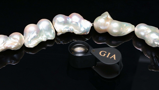
GIA’s Hong Kong laboratory recently examined a necklace consisting of 24 white baroque-shaped nacreous pearls ranging in size from 21.92 × 15.24 mm to 24.95 × 15.12 mm (figure 1). Their external appearance, high luster, and strong orient hinted at a freshwater origin. Detailed examination revealed additional features of interest.
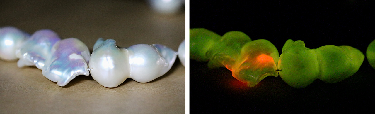
While their appearance was indicative of freshwater pearls, energy-dispersive X-ray fluorescence analysis was performed on a random selection to confirm their formation environment. The high manganese concentrations of 751–1728 ppm were consistent with freshwater pearls. In keeping with the reactions seen in freshwater pearls with such levels of Mn, the samples exhibited strong green fluorescence when exposed to X-rays. However, one in particular displayed moderate to strong reddish orange fluorescence on some areas of its surface (figure 2), associated with whitish non-nacreous regions on the pearl. Similar reddish fluorescence reactions had previously been observed around damaged areas of nacre on pearls examined in GIA’s New York laboratory (Summer 2013 Lab Notes, pp. 113–114). The exact reason for this reaction, which GIA gemologists have seen in cultured and natural freshwater pearls from time to time, is unknown.

Eleven of these pearls had a shape resembling a dumbbell or peanut shell, indicating the possible presence of two nuclei in each. Real-time microradiography confirmed the presence of two round bead nuclei in these pearls (figure 3). The remaining 13 contained only a single round bead, consistent with the majority of bead-cultured pearls on the market. In addition, a network of tubules was observed within most of the bead nuclei. Such tubules resembled those observed in saltwater shells, which result from the burrowing actions of parasites. Thus, the bead nuclei were likely fashioned from saltwater shells rather than the usual freshwater nuclei employed. Nonetheless, it was impossible to determine the nature of the shell beads without destructive testing. Microradiography examination proved the bead nuclei were not pre-drilled, a characteristic often observed in freshwater nuclei used to culture freshwater pearls (H.A. Hänni, “Ming pearls: A new type of cultured pearl from China,” Journal of the Gemmological Association of Hong Kong, Vol. 32, 2011, pp. 23–25) and typically encountered by GIA during testing. While pearls cultured with two bead nuclei are relatively uncommon, their existence has been acknowledged in the literature (Fall 1993 Lab Notes, pp. 202–203). The twin bead structure clearly influenced the appearance of these pearls and resulted in some pleasing baroque shapes.

Finally, upon closer examination, a “glittery” grainy material was observed on the pits and blemishes of several pearls (figure 4). This resembled the essence d’orient applied on the surface of some imitation pearls. Raman analysis of this material yielded a number of peaks (e.g., 634–639 cm–1 and 1604 cm–1) that matched, to some degree, the peaks of the filling material described in an earlier reference (Winter 2017 GNI, pp. 482–484).
While the GIA laboratory examines pearl strands on a daily basis, we seldom encounter one submission possessing several interesting features such as the twin bead structure, reddish orange optical X-ray fluorescence, and filled surface blemishes. It is important for gemologists to always stay alert to the surprises they might find when examining pearls.


