Colored Bands in CVD-Grown Diamond


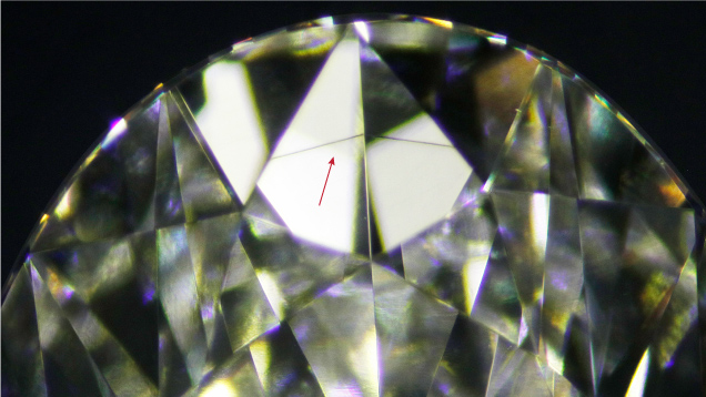
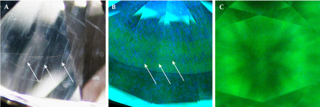
The Surat laboratory recently examined a 3.14 ct F-color oval brilliant diamond grown by chemical vapor deposition (CVD). The diamond featured a single dark brown band measuring ~2.2 mm in length that resembled graining in natural diamond (figure 1). The band was visible under the microscope as well as with a 10× loupe. The clarity grade was VVS2 based on this colored band, which was visible through multiple bezels and affected the transparency at that location. Through the pavilion, parallel whitish bands were also observed (figure 2A).

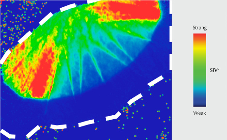
The subtle banding seen in this diamond differed from a cloud of graphite inclusions at a growth interface previously reported in a CVD-grown diamond (Summer 2023 Lab Notes, pp. 213–214). The fluorescence image collected by the DiamondView revealed a layered growth structure that did not coincide with the color banding, indicating a start-stop cycling growth process typical of CVD synthesis (figure 2B). Deep UV fluorescence with green and blue coloration as well as strong green phosphorescence seen in the DiamondView image (figure 2C) indicated high-pressure, high-temperature treatment. The SiV– defect at 736.6 and 736.9 nm, a common feature of CVD laboratory-grown diamond and only rarely seen in natural diamond, was observed in photoluminescence (PL) spectra using 457, 514, and 633 nm laser excitation. PL mapping (figure 3) revealed that the concentration of SiV– was higher near the culet of the pavilion and dramatically lower near the table.
GIA has documented growth remnants in thousands of CVD-grown diamonds. But with a multitude of manufacturers, recipes, and treatments, a wide variety of clarity characteristics are encountered, including the unusual color band observed here.


