CVD-Grown Diamond with Few Diagnostic Features

Diamonds grown by chemical vapor deposition (CVD) have evolved rapidly in the last two decades. However, the vast majority submitted to GIA for reports still show several diagnostic features such as the silicon vacancy or SiV– defect (a doublet at 736.6 and 736.9 nm) and/or the 468 nm peak when analyzed by photoluminescence (PL) spectroscopy. Additionally, most CVD diamonds have distinctive fluorescence images when analyzed by deep UV with the DiamondView instrument.

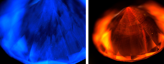
Therefore, it was interesting to analyze a 1.40 ct E-color, VVS2 type IIa diamond submitted for a laboratory-grown diamond report. Initial PL data collection did not show either the SiV– center or the 468 nm peak. The features observed were a 3H peak at 503.5 nm using 455 nm excitation and relatively weak nitrogen vacancy (NV) centers at 575 (NV0) and 637 (NV–) nm using 514 nm excitation (figure 1, left). The stone showed weak anomalous birefringence indicating strain; while this observation is helpful in confirming whether a diamond is grown by high-temperature, high-pressure (HPHT) methods, such birefringence cannot distinguish between natural and CVD origins. The deep-UV fluorescence image showed only deep blue coloration, which appeared generally comparable with the vast majority of natural type IIa diamonds (figure 2, left). Therefore, many of the standard markers of CVD origin were not present even when examined using advanced methods. Careful analysis of the fluorescence image revealed some distinct differences from the majority of natural diamonds, namely the patchiness of the fluorescence with some inert regions.
Many models of the DiamondView instrument include a series of filters that permit fluorescence imaging in which portions of the visible spectrum are blocked out. Using these filters, the blue portion of the visible spectrum was blocked in order to observe weaker underlying fluorescence features. With the orange long-pass filter blocking wavelengths less than 550 nm, NV-related fluorescence clearly showed the striations indicative of CVD growth (figure 2, right). Also, several locations of the diamond were analyzed with PL spectroscopy to determine if either the 468 nm center or the SiV– could be detected. Only at the culet was a small 468 nm feature observed (figure 1, right).
The author concluded that this was a CVD-grown diamond with no evidence of post-growth treatment. This example is a noteworthy indicator that as CVD growth methods improve, telltale defects are being minimized or eliminated entirely. Careful examination of all data, both spectroscopic and fluorescence imaging, is needed to provide accurate origins for diamond.


