Mabe Cultivation in Mayotte

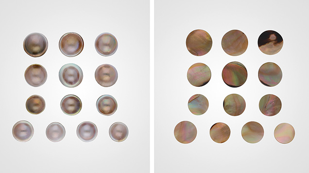
The Laboratoire Français de Gemmologie (LFG) received some mabe samples (figure 1) from Antoine Ganne of the Mayotte-based company Nuru Kombe for gemological characterization. According to the company, these samples were cultivated in Pteria penguin bivalves in a lagoon off the coast of Mayotte. This French territory is situated in the Indian Ocean between northwestern Madagascar and northeastern Mozambique, with one main island and several islets.
Mabe is the Japanese term designating an assembled cultured blister, traditionally from Pteria penguin but nowadays from other mollusks as well (CIBJO Pearl Commission, The Pearl Book, CIBJO, Milan, 2020, 79 pp.). Mabe is made of purpose-grown cultured blisters that have been cut from their shell. The original bead upon which they grew is removed, and the cavity is filled with various synthetic materials. It is then backed by a layer of shell, with the assemblage held together by an adhesive.
The studied samples ranged from light brown to brown (some of a “golden” color) with pronounced secondary colors such as blue, green, and red (again, see figure 1). Under the microscope, all samples presented the characteristic nacreous structures associated with overlapping layers of aragonite. The curved and flat sides looked very similar, suggesting they originated from the same mollusk species. The samples reacted purple to red under a long-wave UV lamp and remained inert under short-wave UV lamp excitation. Those with a more pronounced brown color exhibited a stronger intense luminescence. A similar luminescence reaction was observed in cultured pearls from Pteria sterna from Mexico (L. Kiefert et al., “Cultured pearls from the Gulf of California, Mexico,” Spring 2004 G&G, pp. 26–38). This response is due to a type of porphyrin (a tetrapyrrole with a cyclic structure) that also plays an important role in the samples’ coloration.

Raman analysis using a 514 nm laser revealed bands associated with aragonite along with a series of bands from 800 to 1600 cm–1 characteristic of porphyrins (S. Karampelas et al., “Raman spectroscopy of natural and cultured pearls and pearl producing mollusc shells,” Journal of Raman Spectroscopy, Vol. 51, No. 9, 2020, pp. 1813–1821). Photoluminescence spectra obtained using a Raman spectrometer and the same laser excitation presented bands in the orange and red part of the electromagnetic spectrum at about 620, 650, and 680 nm (figure 2). The relative intensity of these bands can vary. Luminescence spectra using a xenon lamp with 365 nm excitation presented similar bands with additional bands at about 435, 465, and 525 nm. The latter bands are possibly linked to the nacre of the samples, while the triplet of bands at 620, 650, and 680 nm has been attributed to a uroporphyrin (Y. Iwahashi and S. Akamatsu, “Porphyrin pigment in black-lip pearls and its application to pearl identification,” Fisheries Science, Vol. 60, No. 1, 1994, pp. 69–71).

The UV-Vis reflectance spectra of the samples featured a series of bands with various relative intensities at about 380, 400, 460, and 495 nm, along with a band at about 280 nm (figure 3). The latter band is linked to organic matter and the nacreous part of the samples, and the series is possibly linked to porphyrins (Kiefert et al., 2009).

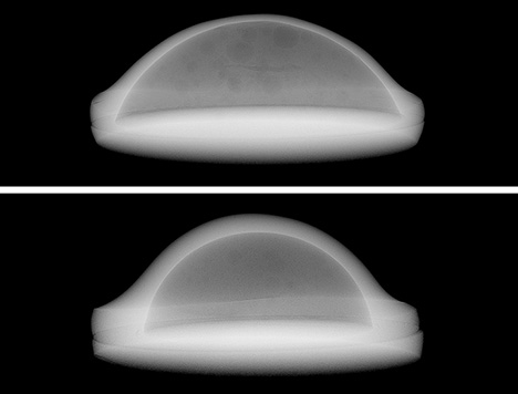
All samples were inert under X-ray and EDXRF analysis, with an Mn content below the detection limit and Sr contents ranging from 3180 to 5280 ppm, indicating saltwater origin. X-ray microradiographs (figure 4) showed lighter tones at the rim, indicating a higher-density material such as calcium carbonate (usually aragonite) in the case of natural and cultured pearls, while the darker tones in the core indicated lower-density material such as organic matter and resin (S. Karampelas et al., “Real-time microradiography of pearls: A comparison between detectors,” Winter 2017 G&G, pp. 452–456). The samples, which could be called “mabe” or “assembled cultured blister” according to the current CIBJO standards, had a nacre thickness from 0.4 to 1 mm (again, see figure 4).
Mabe from Mayotte has only appeared recently in the market. The exact supply is still unknown, and other similar products are cultivated in other parts of the world, including Southeast Asia, Australia, and some Pacific island nations (P.C. Southgate and J.S. Lucas, The Pearl Oyster, 2008, Elsevier, Amsterdam). Nevertheless, this material could help develop the economy of Mayotte, where all the operations take place and jewelry pieces with a French touch are locally made.


