Black Non-Nacreous Pearl Imitations Made of Beads Cut from Shell
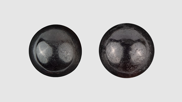
DANAT (Bahrain Institute for Pearls & Gemstones) recently received a 5.04 ct bead (9.22 × 9.15 × 8.67 mm) and a 5.45 ct bead (9.51 × 9.44 × 8.69 mm), both of near-round shape (figure 1). Under the microscope, both beads presented columnar structures in some areas, similar to those observed in some non-nacreous pearls (N. Sturman et al., “Observations on pearls reportedly from the Pinnidae family (pen pearls),” Fall 2014 G&G, pp. 202–215), as well as elongated structures due to fibers reaching the surface (figure 2). Raman spectra with a 514 nm green laser were difficult to acquire, as they revealed high luminescence possibly linked to their high organic matter content; however, there was enough Raman signal to be able to identify calcite and aragonite. The calcium carbonate identification was confirmed by acquiring FTIR reflectance spectra. In the Raman spectra, weak bands linked to polyenic pigments were also observed.

Chemical analysis of the beads using energy-dispersive X-ray fluorescence (EDXRF) did not show any measurable manganese. Strontium was above 800 ppmw, and they remained inert under X-ray imaging, similar to saltwater pearls. Digital X-ray microradiographs and micro-CT did not show any internal structures.


Under long-wave and short-wave ultraviolet light, zoned/banded/layered fluorescence was observed (figure 3). Using transmitted fiber-optic illumination, banded structures and fibrous structures were visible with the unaided eye (figure 4). These characteristics were similar to those observed in imitation pearls made of non-nacreous shell material. Most of the imitation pearls described previously had a light color or were artificially dyed. The beads examined in this study, though, presented no evidence of any color treatment under the microscope or using EDXRF and Raman spectroscopy. The “zoned” fluorescence reaction under UV light was linked with different structures and not with the artificial colorants.
Large white beads with fibrous aragonite supposedly cut from bivalves from Tridacna species, which contain thick inner layers, have previously been used to imitate pearls (Summer 2004 GNI, p. 178; Summer 2006 Lab Notes, pp. 166–167; M. Krzemnicki and L. Cartier, “Fake pearls made from Tridacna gigas shells,” Journal of Gemmology, Vol. 35, No. 5, 2017, pp. 434–429). These beads were sometimes dyed to alter their coloration. A salmon-colored bead of about 10 mm, supposedly cut from the inner part of Lobatus gigas, was used to imitate coral (E. Disner and F. Notari, “Gastropod shell beads disguised in a coral necklace,” Journal of Gemmology, Vol. 34, No. 7, 2015, pp. 572–574). The beads we examined were of natural black color, and we are still looking for a mollusk that might have a non-nacreous inner shell layer where beads of that size could be cut.
Acknowledgments: The authors would like to extend their appreciation to Mr. Saud Bin Rajab (Manama, Bahrain) for allowing us to publish the study on these beads.


