True Colors of “Dalmatian Jasper”

ABSTRACT
“Dalmatian jasper,” also known as “dalmatian stone,” is a popular decorative gemstone with a unique appearance resembling the spotted coloration of the Dalmatian dog breed. Our research revealed that this gemstone is a peralkaline rock of uncertain provenance. It consists predominantly of feldspars (mesoperthite), quartz, alkali amphiboles, and lesser amounts of hematite and epidote. Black spots found in the examined rock’s mass were recognized as arfvedsonite. The authors recommend the use of the term “dalmatian stone” rather than “dalmatian jasper,” since the material does not meet the gemological definition of jasper.
INTRODUCTION
Jasper (from the ancient Greek for “spotted stone”) has a variety of colors, patterns, and textures that make it a very popular gemstone worn in jewelry and sought after by collectors and geologists. Unfortunately, the term “jasper” has become commercialized and misused by many sellers. In addition, the nomenclature and classification of jaspers are not well coordinated in gemology and petrology (Kostov, 2010). Manutchehr-Danai (2010) considered jasper a dense, translucent to opaque impure variety of chalcedony or chert. Götze (2010) defined it as a mixture of microcrystalline silica with iron oxide and illite, while Kievlenko (1980) classified it as a metamorphic or metasomatic material. Hence, the jasper group is characterized by sedimentary, metamorphic, or igneous rocks. Examples include, respectively, radiolarites such as mookaite jasper (Campos-Venuti, 2012), quartzites such as Sioux Falls jasper (Dietrich, 2009), and rhyolites such as orbicular jasper and picture jasper (Żaba, 2010; Polk, 2012). In general, all jasper types have specific physical, chemical, and aesthetic properties that make them a valuable material for jewelry. Except for silica, most also contain some admixtures such as iron oxides/hydroxides, feldspars, and epidotes (Żaba, 2010).
In some cases, more detailed mineralogical and petrological studies are necessary to determine the particular type of material. “Dalmatian jasper,” also known as “dalmatian stone,” is a misnomer if we consider the gemological definition of jasper or even other spotted stones mentioned in the literature (some of which were presented in the previous paragraph). Dalmatian stone consists of a white to grayish rock mass devoid of larger phenocrysts, with small crystals of quartz visible to the unaided eye. The rock mass strongly contrasts with the black to greenish spherical assemblages of minerals that are responsible for the unique dalmatian pattern. The diameter of these black spots usually does not exceed 4 mm. The surface of the rock is sometimes covered by red to brown iron oxide/hydroxide minerals.
Published information on dalmatian stone and its mineralogical composition is quite meager. Based only on microscopic observations, Bruder (2006) recognized dalmatian stone as an aplite, consisting predominantly of quartz, feldspars (albite and microcline), and Fe-riebeckite. On the other hand, Campos-Venuti (2012) considered it a variety of devitrified rhyolite and classified it as “rhyolitic jasper.” In contrast, some informational websites on various gemstones (e.g., www.healing-crystals-for-you.com/dalmatian-jasper.html) indicate that the black spots in dalmatian stone are composed of tourmaline-group minerals. Moreover, the source of this gemstone has not yet been specified (Dietrich, 2009).
The aim of this work is to determine the detailed mineralogical composition of dalmatian stone, classify it properly, and discuss potential locations of the rock’s outcrops proposed by distributors of the material. To accomplish this we applied microscopic observations in transmitted light, scanning electron microscopy, Raman microspectroscopy, and chemical analysis.
MATERIALS AND METHODS
Three samples of dalmatian stone, including a rough sample, bracelet, and pendant from an unknown locality, were purchased during the International Exhibition and Trade Fair of Minerals, Fossils, and Jewellery at the Krakow University of Economics in Poland. All samples were marked by high hardness and could take a polish. They revealed a spotted texture typical of dalmatian stone (black spots within white to grayish rock matrix) and were poorly altered by weathering processes. The raw specimen was block-shaped (1.5 × 1.5 × 1 cm) and had a rugged surface (figure 1, left). The bracelet consisted of polished beads with waxy luster measuring 1 cm in diameter, while the pendant had an oval shape and measured about 3 cm in length (figure 1, right). These samples were analyzed using polarizing microscopy, scanning electron microscopy, Raman microspectroscopy, and chemical analysis via inductively coupled plasma–emission spectrometry (ICP-ES) and inductively coupled plasma–mass spectrometry (ICP-MS).
A thin section of the dalmatian stone rough was examined with an Olympus BX51 polarizing microscope with a magnification range from 40× to 400×. Photos were taken using an Olympus DP12 digital camera with Analysis software. Backscattered electron (BSE) images of the polished section were obtained using an FEI Quanta 200 FEG scanning electron microscope equipped with an energy-dispersive spectroscopy (EDS) detector. The system was operated at 25 kV accelerating voltage in a high-vacuum mode.
Raman spectra of black spots seen with the unaided eye were recorded using a Thermo Scientific DXR Raman microscope equipped with 100×, 50×, and 10× magnification objectives, operated in confocal mode and in a backscatter geometry. The samples were excited with a 532 nm laser. The laser focus diameter was approximately 1–2 μm. Acquisition time was 20 seconds. Almost no sample preparation was performed. The polished section of dalmatian stone was cleaned carefully with distilled water and acetone before measurements were made to ensure that it was not contaminated. The spectra were acquired up to 2500 cm–1 using Omnic Specta software. Phase identification was performed by CrystalSleuth software in conjunction with the RRUFF database (http://rruff.info). The assignment of particular Raman-active bands was made using data for alkali amphiboles (especially riebeckite) published by Apopei and Buzgar (2010).The whole-rock analyses were performed at Bureau Veritas Minerals Laboratories Ltd. in Vancouver, Canada. The abundances of major oxides and trace elements were determined using ICP-ES and ICP-MS. The sample was melted and dissolved by lithium borate flux. The results of representative whole-rock analyses are presented in table 1.
| TABLE 1. Whole-rock major and trace-element analysis of dalmatian stone. | |||||||
| Major element (wt.%) | Trace element (ppmw) | ||||||
| SiO2 | 73.66 | Be | 16.00 | Zr | 2072.10 | Nd | 153.50 |
| TiO2 | 0.21 | Rb | 374.30 | Nb | 156.50 | Sm | 30.17 |
| Al2O3 | 10.74 | Sr | 42.30 | Hf | 53.40 | Eu | 0.38 |
| FeOatot | 4.09 | Cs | 7.60 | Ta | 9.40 | Gd | 28.76 |
| MnO | 0.07 | Ba | 144.00 | Th | 36.60 | Tb | 4.90 |
| MgO | 0.08 | V | 17.00 | U | 14.20 | Dy | 30.63 |
| CaO | 0.48 | Co | 0.50 | Pb | 15.60 | Ho | 5.99 |
| Na2O | 4.40 | Ni | 2.10 | Ga | 41.90 | Er | 19.33 |
| K2O | 4.65 | Cu | 1.70 | La | 176.50 | Tm | 2.88 |
| P2O5 | 0.01 | Zn | 48.00 | Ce | 361.40 | Yb | 18.92 |
| LOIc | 1.10 | Y | 163.70 | Pr | 42.95 | Lu | 2.88 |
| Total | 99.49 | ||||||
| PId | 1.14 | ||||||
|
aFeOtot = total Fe as Fe2O3
bbdl = below detection limit cLOI = loss on ignition dPI = peralkaline index (Na2O + K2O/Al2O3) |
|||||||
RESULTS
Microscopic Characteristics. Dalmatian stone is described as homogeneous, massive, and unfoliated rock. The rock matrix of the sample examined in this study consisted predominantly of quartz, feldspars (mostly mesoperthite), and subordinate alkali amphiboles (arfvedsonite). Epidote-group minerals as well as hematite and goethite formed the secondary phases. Quartz crystals in the thin section reached sizes up to 0.3 mm (figure 2). These crystals appeared to be sharp-edged because they were often partially overgrown with smaller feldspar crystals. At the contact with alkali amphiboles, some of the quartz crystals exhibited an oval shape.
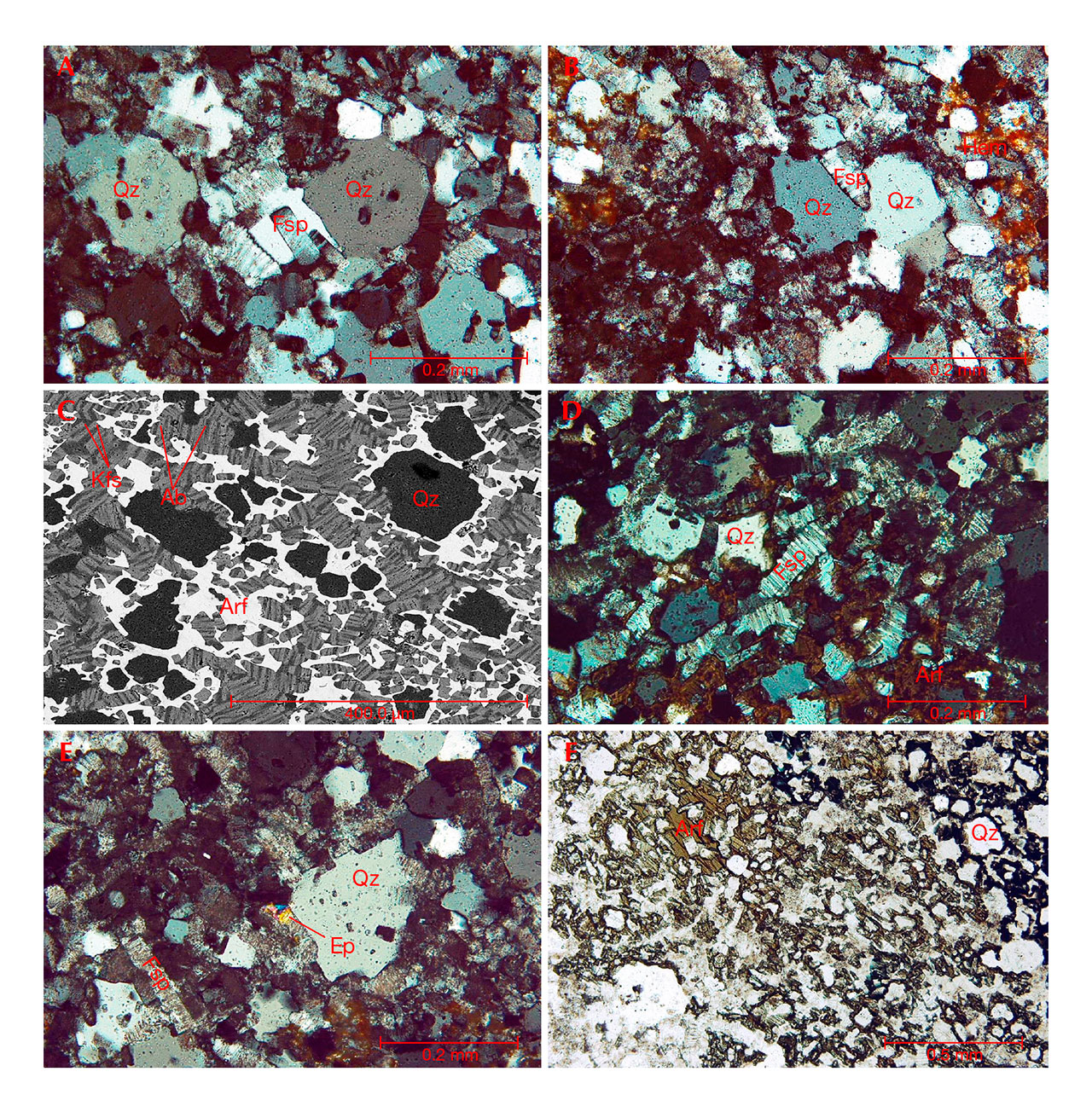
Feldspars were represented mostly by mesoperthites (intergrowths of K-feldspar with Na-feldspar) up to 0.2 mm in size (figures 2C and 2D). Their crystals were pigmented with hematite and poorly altered. Some were partially enclosed between larger quartz crystals (figures 2A and 2B). Locally, feldspar crystals exhibited Carlsbad twinning and were bent and dislocated, likely resulting from the deformation of magmatic rock components that were not completely solidified.
Alkali amphiboles were represented by anhedral crystals that filled the interstitial spaces between quartz and feldspars (figures 2C and 2F). They usually formed radial-like aggregates. Typical pleochroic colors (X = greenish blue, Y = brownish green, Z = black) identified the alkali amphiboles as arfvedsonite. They were partially altered into iron oxides or hydroxides (hematite and goethite) due to weathering processes.
Minerals from the epidote group formed small crystals or granular aggregates up to 0.05 mm (figure 2E). They were pale green and exhibited second-order interference colors (blue, yellow, and pink) within a single crystal.
Raman Spectroscopy of Alkali Amphiboles. The Raman spectra of mafic minerals revealed the presence of arfvedsonite and poorly crystalline hematite (figure 3). The most distinct band of arfvedsonite is located at 675 cm–1 and may be ascribed to ν1/νs (symmetric stretching vibrations) of the Si-Ob-Si bridges (Apopei and Buzgar, 2010). Arfvedsonite has a relatively complex chemical composition, and concentrations of K, Mg, Mn, or Fe in its structure may have a huge influence on peak location or intensity. Most of the Raman spectra recorded in this study exhibited high fluorescence related to the significant amounts of Fe in the structure of alkali amphiboles. Location and assignment of the bands are listed in table 2.
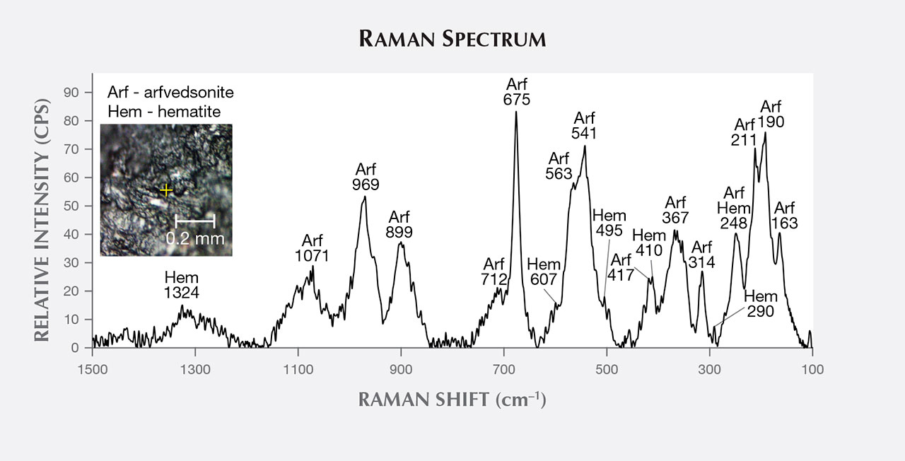
| TABLE 2. Raman band positions (cm–1) and their assignments for arfvedsonite in the1500–100 cm–1 spectral region. | ||
| Present study | Lafuente et al. (2015) | Assignment |
| 163 | 170 | Lattice mode |
| 190 | 199 | |
| 211 | 204 | |
| 248 | 240 | |
| 314 | 317 | M-O |
| 367 | 357 | |
| 417 | 418 | |
| 541 | 533 | Deformation modes of Si4O11 |
| 563 | 558 | |
| 675 | 675 | νs of Si-Ob-Si (ν1) |
| 712 | 721 | |
| 899 | 879 | νs of O-Si-O |
| 969 | 981 | νas of O-Si-O |
| 1071 | 1094 | νas of Si-Ob-Si |
|
νs = symmetric stretching
νas = asymmetric stretching M-O = vibration modes from the interactions between the cation and oxygen Ob = bridging oxygen O = non-bridging oxygen |
||
The broad peak located at 1324 cm–1 is diagnostic for disordered hematite and should be assigned to a second-order 2LO mode with 2Eu symmetry due to defects in the hematite lattice (Marshall and Marshall, 2013). Hematite bands at 225, 247, 412, 498, and 613 cm–1, which are specific to this mineral according to Legodi and de Waal (2007), overlap with arfvedsonite bands. Consequently, they are hard to distinguish. In addition, the intensity of the arfvedsonite peaks in these spectral regions is quite amplified. The location and assignment of particular bands of hematite are listed in table 3. The presence of this mineral in the measured sample probably originated from the weathering of arfvedsonite.
| TABLE 3. Raman band positions and their assignments for poorly crystalline hematite in the1500–100 cm–1 spectral region. | ||
| Present study (cm–1) |
Previous studiesa (cm–1) |
Assignment |
| 211 | 225 | A1g |
| 248 | 247 | Eg |
| 290 | 293 | Eg |
| 410 | 412 | Eg |
| 495 | 498 | A1g |
| 607 | 613 | Eg |
| 1324 | 1320 | 2Eu |
|
aSources: Legodi and de Waal (2007); Marshall and Marshall (2013)
Eg = Fe-O symmetric bending A1g = Fe-O symmetric stretching 2Eu = 2LO mode |
||
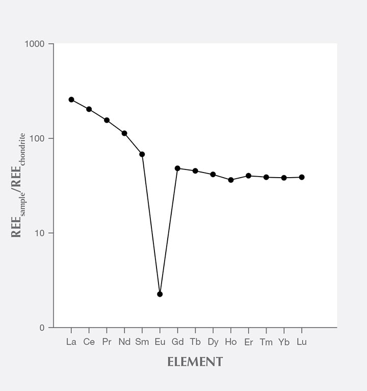
Geochemistry. The major chemical constituent of the rock in this study was silica (73.66 wt.% SiO2). The amount of Al2O3 was relatively low at almost 11 wt.% (table 1). The rock was slightly enriched in alkalis: Na2O (4.40 wt.%) and K2O (4.65 wt.%). In contrast, it was depleted in MgO and CaO; their contents did not exceed 0.5 wt.% (table 1). The analyzed sample was enriched in light rare-earth elements (LREEs): [La/Sm]N>1, [Sm/Yb]N>1, and [La/Yb]N=6.69. It contained relatively high amounts of high-field-strength elements (HFSE) such as zirconium and niobium (2072.1 ppm and 156.5 ppm, respectively). The calculated peralkaline index (Na2O+K2O/Al2O3 molecular ratio) for this rock was 1.14. The normative composition, calculated using a common algorithm to estimate the standard mineral assemblages for igneous rocks, revealed the presence of quartz (30.26 wt.%), plagioclase (29.67 wt.%; anorthite content 0 wt.%), orthoclase (27.91 wt.%), acmite (6.94 wt.%), diopside (2.11 wt.%), hypersthene (2.07 wt.%), zircon (0.42 wt.%), ilmenite (0.4 wt.%), sodium metasilicate (0.04 wt.%), and apatite (0.02 wt.%).
DISCUSSION AND CONCLUSIONS
Our dalmatian stone sample had a specific mineral assemblage that included quartz, feldspar mesoperthites, and alkali amphiboles (arfvedsonite). It contained acmite in its normative composition and had a peralkaline index higher than 1. As a result, we classify dalmatian stone as a peralkaline rock. Textural features were not distinctive enough to determine whether the rock had an intrusive or extrusive origin. The dalmatian stone specimen examined in our work may represent peralkaline microgranite or aplite as well as peralkaline rhyolite, since the composition of these rocks may be identical. Intrusive origin is supported by the fact that the samples examined in this study do not exhibit the porphyric texture or the glassy or cryptocrystalline groundmass that together would indicate a relatively fast cooling. On the other hand, quartz crystals may be treated as phenocrysts because they are slightly larger than other crystals, which could support an extrusive origin. Moreover, interstitial arfvedsonite, in the same form as in our photomicrographs, has been reported in comenditic rhyolite (see www.alexstrekeisen.it/english/vulc/comendite.php).
Peralkaline rocks are generally abundant in continental rift settings (Mbowou et al., 2012; Shao et al., 2015). They are considered end members of the sodic (Atlantic) magma differentiation series (Majerowicz and Wierzchołowski, 1990). The most distinct feature of these rocks is depletion of Al2O3 and enrichment in Na2O and K2O. The depletion of Al at the late stage of melt crystallization is compensated by Fe. As a result, Fe-bearing alkali amphiboles (e.g., riebeckite and arfvedsonite) could have crystallized at that time. The relatively small amounts of Al in peralkaline rocks probably originate from the “plagioclase effect” that happened with removal of Ca-rich plagioclase (CaAl2Si2O8) at an early stage of magma evolution. Such Ca-rich plagioclases have twice as much alumina as alkali feldspars, so their crystallization depletes Al from magma and causes the relative enrichment of Na and K (Mbowou et al., 2012; Shao et al., 2015). The primitive mantle rare-earth element (REE) pattern of dalmatian stone (figure 4) is similar to that reported in peralkaline rocks (e.g., Mbowou et al., 2012) and reveals the strong depletion of europium (also known as a negative europium anomaly). This is due to the fact that plagioclases, which were not found in this study, tend to be enriched in europium.
The microscopic and chemical analyses were used to determine not only the classification of dalmatian stone but also its petrogenesis. Feldspar and quartz probably represent the earliest generations of minerals. The nucleation of alkali amphiboles occurred at the late stage of dalmatian stone’s crystallization, forming as anhedral crystals filling the interstitial space between quartz and feldspars. Sodium plagioclases do not occur as individual, discrete crystals but form exsolution lamellae in K-feldspars. Thus, dalmatian stone may be classified as hypersolvus rock, which contains a single feldspar (sometimes with exsolution lamellae) and forms under low H2O pressures and relatively high temperatures (e.g., Klein and Philpotts, 2013). Epidote-group minerals probably formed during the post-magmatic stage. They also could have crystallized from fluids originating from surrounding rocks (Vlach, 2012).
The provenance of the rock from this study and Dietrich (2009) remains unknown. One possible source of dalmatian stone indicated by gemstone dealers is the Mexican state of Chihuahua. This seems very probable because of the occurrence of Burro Mesa riebeckite rhyolite at Big Bend National Park in the neighboring U.S. state of Texas (reported by Maxwell et al., 1967). These rocks were described as highly siliceous, medium-grained gray rhyolite with quartz phenocrysts in a riebeckite matrix. Nevertheless, further investigations are necessary to prove this hypothesis. It should also be mentioned that dalmatian stone may resemble Capo Bianco aplite from the Mediterranean island of Elba. This rock consists of characteristic black tourmaline nodules that contrast strongly with its white groundmass (see Perugini and Poli, 2007).
Dalmatian stone has a unique pattern, but its mineral composition can be identified with advanced and non-destructive methods such as Raman spectroscopy or SEM-EDS. As discussed above, “dalmatian jasper” should be considered a trade name for peralkaline rock (microgranite, aplite, or rhyolite). Eventually, it may be classified—together with such rocks as orbicular jasper or picture jasper—in a group of so-called jasper-like intrusive or effusive rocks with feldspar-quartz composition (see Putolova et al., 1989). To distinguish dalmatian stone from jaspers and jasper-like rocks, it is necessary to make macroscopic observations supported by Raman microspectroscopy.


