A New Deposit of Gem-Quality Grandidierite in Madagascar
ABSTRACT
A new deposit of grandidierite, considered one of the world’s rarest gems, has been discovered in southern Madagascar. The new deposit is outside the town of Tranomaro, near the original locality of Andrahomana. It occurs in the form of strong bluish green to greenish blue translucent to transparent crystals measuring up to 15 × 7 × 3 cm. Grandidierite is the magnesium end member in the solid-solution series with ominelite as the iron end member. The studied samples have a very low Fe/(Mg + Fe) ratio. This confirms that the Tranomaro deposit, together with Johnsburg in New York State, provides the purest grandidierite ever found. The crystals host inclusions of Cl-apatite, zircon, and monazite. The paragenesis also includes plagioclase, phlogopite, enstatite, diopside, dravite, and sapphirine (locally as gem-quality crystals). Transparent crystals have been faceted, yielding small but eye-clean jewelry-quality gems.
INTRODUCTION
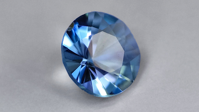
Named after French naturalist Alfred Grandidier (1836–1912), grandidierite is an extremely rare orthorhombic Mg-Fe aluminous borosilicate with the formula (Mg,Fe)Al3(BO3)(SiO4)O2 (Lacroix, 1902, 1922a,b). The material was first discovered at the cliffs of Andrahomana, on the southern coast of Madagascar. Grandidierite is described as a bluish green to greenish blue mineral (figure 1); the blue component increases with Fe content. It is transparent to translucent with a pale yellow to colorless, greenish blue, and blue trichroism. The larger elongate euhedral orthorhombic crystals, which may measure up to 8 cm, are often strongly corroded.
Since its discovery, grandidierite has been found as a rare accessory mineral in aluminous boron-rich pegmatite; in aplite, gneiss, and crystalline rock associated with charnockite; and in rock subjected to local high-temperature, low-pressure metamorphism (contact aureoles and xenoliths). In addition to Madagascar, it has been reported from New Zealand (Black, 1970), Norway (Krogh, 1975), Suriname (de Roever and Kieft, 1976), Algeria (Fabriès et al., 1976), Italy (van Bergen, 1980), Malawi (Haslam, 1980), India (Grew, 1983), the United States (Rowley, 1987; Grew et al., 1991), Canada (Lonker, 1988), Antarctica (Carson et al., 1995), the Czech Republic (Cempírek et al., 2010), and other localities. Yet gem-quality grandidierite is extremely rare. Facetable gem material larger than a millimeter has only been found in Madagascar and Sri Lanka; the latter is the source of a clean faceted specimen weighing 0.29 ct (Schmetzer et al., 2003).
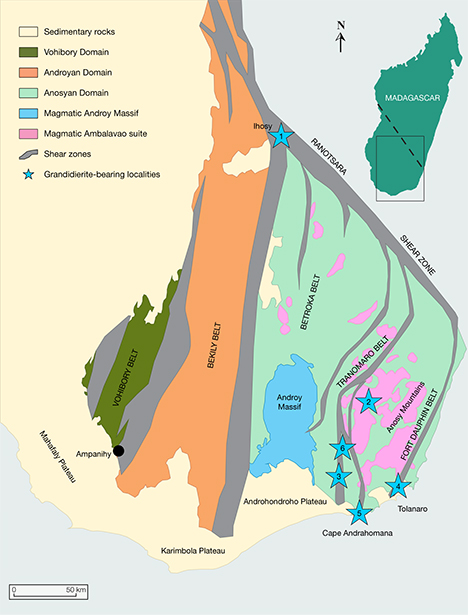
The type locality described by Lacroix (1902) was visited in 1960 by C. Mignot, who was unable to find additional grandidierite at the site (Béhier, 1960a,b), as the small pegmatite was depleted. Since then, grandidierite has been reported in a few other localities in southern Madagascar that have also since been depleted. The present study focuses on a new deposit discovered in May 2014 near Tranomaro, close to the original locality of Andrahomana (figure 2). The aim of this study is to determine the material’s properties and to evaluate its gem potential.
GEOLOGICAL BACKGROUND
The crystalline bedrock of Madagascar is described in terms of six geodynamic blocks that define the constituent elements of the Precambrian Shield; these are, from southwest to northeast, the Vohibory, Androyan-Anosyan, Ikalamavony, Antananarivo, Antongil-Masora, and Bemarivo domains. As the result of a major five-year (2007–2012) study funded by the World Bank, the Precambrian geology of Madagascar has been reinterpreted. The Antongil-Masora and Antananarivo domains are now considered part of an even larger craton—the Greater Dharwar craton—that encompassed part of present-day India and Madagascar and was consolidated approximately 2.45–2.5 billion years ago (Tucker et al., 2012). The Ranotsara shear zone marks the approximate boundary between the Archean crust to the north and a more recent basement to the south. The Androyen-Anosyen domain of southern Madagascar is indeed a continental terrane of Paleoproterozoic age (1.8–2.0 billion years ago). It was accreted to the Vohibory domain, an oceanic terrane of early Neoproterozoic age, in Cryogenian time (about 0.62–0.64 billion years ago). A first deformation (600–650 Ma) gave rise to recumbent isoclinal folds and a regional shallow-dipping foliation, with a second deformation (500–530 Ma) producing steep north-south-trending shear zones that separate five gneissic tectonic belts. The last episode of Madagascar’s Neoproterozoic geodynamic evolution is marked by the alkaline magmatism of the Ambalavao suite, which forms major batholitic intrusions (up to 1400 km2). Numerous pegmatitic vein intrusions associated with this episode are directly related to gem and rare earth element (REE) mineralization. A final magmatic activity during the Cretaceous period is expressed by the megavolcanic Androy Massif.
A grandidierite-bearing pegmatite is found in the Anosyan domain (again, see figure 2). At Cape Andrahomana, where grandidierite was first described, it was associated with quartz, microcline, red almandine, pleonaste, andalusite, and biotite in a pegmatite and an aplite (Lacroix, 1922a,b). Grandidierite was also found 30 km to the north, near Vohibola, in association with serendibite and sinhalite in a diopsidite-bearing paragneiss (Razakamanana et al., 2010). It was associated with quartz in the Nampoana quarry near Tolanaro (Farges, 2001) and at Sakatelo, Ampamatoa, Marotrana, and Sahakondra, near Esira (McKie, 1965; Farges, 2001). It was also found in association with serendibite and tourmaline in anatectic gneiss near Ihosy (Nicollet, 1990). These reports relate only microscopic crystals of grandidierite, and the Lacroix finding from a century ago remains the only one of gemological interest. Grandidierite was recently reported from a mine near Tolanaro, with no further information yet (Vertriest et al., 2015).
LOCATION AND MINING
A new deposit of grandidierite was discovered in May 2014 by two of the authors and their mining team, about 15 km from the village of Tranomaro. The area is located in the Amboasary district of southern Madagascar’s Anosy region (again, see figure 2), 60 km northwest of Cape Andrahomana. Access from Tolanaro is via a 60 km paved road west to Amboasary Atsimo, followed by a rough, unpaved 50 km road north to Tranomaro that requires a four-wheel-drive vehicle. Final access from the village of Tranomaro to the deposit is half a day by foot only. Security is a problem because bandits operate throughout the region.
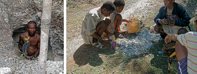
Mining is done by hand due to the locality’s remote location, its limited production, and its irregular, discontinuous veins in opposition to large linear veins. The deposit itself extends over a few acres. The weathered pegmatite is exploited by near-surface artisanal and small-scale mining (figure 3). Using spades and pickaxes, about 12 miners dig holes up to a depth of 15 meters. These open-air corridors cross-cut two grandidierite-bearing veins separated by 30 cm to a few meters. The uppermost tunnel is called “Vein 1,” while the deeper one is “Vein 2.” The rough blue crystals are manually extracted and sorted on-site. The workers carefully remove the valuable mineral specimens to avoid damaging them. Between May 2014 and March 2016, 800 kg of rough specimens were produced. Mining is still in progress as of this writing.
MATERIALS AND METHODS
Using standard gemological methods, we tested a dozen grandidierite crystals from the two veins near Tranomaro for pleochroism, fluorescence reaction, and specific gravity at the French Geological Survey (BRGM) in Orléans, France. After the samples were prepared as polished thin sections, we observed them under a polarizing microscope. The crystals’ chemical homogeneity was investigated with a scanning electron microscope (SEM) on sections that had been carbon coated under vacuum (approximately 20 nm thick). The observations were performed on a Tescan Mira3 XMU field-emission scanning electron microscope (FE-SEM) at 15 kV, and the SEM images were collected using a doped YAG scintillator-type backscattered electron (BSE) detector. Energy-dispersive spectra were obtained by an EDAX TEAM EDS system with an Apollo XPP silicon drift detector.
A Cameca SXFive electron microprobe was used to investigate the grandidierite’s chemical composition, including boron content. The standards were boron nitride (B-Kα), Al2O3 (Al-Kα), orthoclase (K-Kα), MnTiO3 (Mn-Kα, Ti-Kα), andradite (Si-Kα, Ca-Kα), Fe2O3 (Fe-Kα), MgO (Mg-Kα), UO2 (U-Mβ), and ThO2 (Th-Mα). All analyses were performed at an acceleration voltage of 15 kV and a beam current of 20 nA. Count time was 10 s for Mg, Al, Si, K, and Ca; 20 s for Ti; 30 s for Fe and Mn; 60 s for Th and U; and 480 s for B. A ϕρ(Z) method was chosen for data correction. B-Kα intensity was measured in peak area integration mode, while boron intensity was calibrated with a boron nitride reference sample (coated together with the grandidierite samples). The chemical composition is expressed as atoms per formula units (apfu) based on nine atoms of oxygen.
To confirm the mineral identification, powdered samples and thin sections were analyzed by X-ray diffraction (XRD) and Raman microspectroscopy, respectively. The XRD analyses were performed on randomly oriented powders using soda-lime capillaries 0.5 mm in diameter; they were carried out from 6° to 75° 2θ on a Bruker D8 Advance diffractometer with Cu-Kα radiation (λ=1.5418 Å, 40 kV, and 40 mA) and a Lynx-Eye 1D detector, using a step size of 0.04° 2θ and a step time of 1,920 seconds. Bruker’s Diffrac Plus Eva software was chosen for the data analysis. In addition, Raman measurements were performed on polished thin sections with a Renishaw inVia Reflex system coupled to a Leica DM2500 microscope with a 100× objective (numerical aperture = 0.9). The Raman scattering was excited by a 514 nm diode-pumped solid-state (DPSS) laser, and the spectrometer was calibrated using the 520.5 cm–1 line of a silicon standard before each measurement session. Several acquisitions were accumulated to improve the signal-to-noise ratio. Raman spectra of at least 10 spots on each crystal were recorded to ensure the consistency of the data.
RESULTS AND DISCUSSION
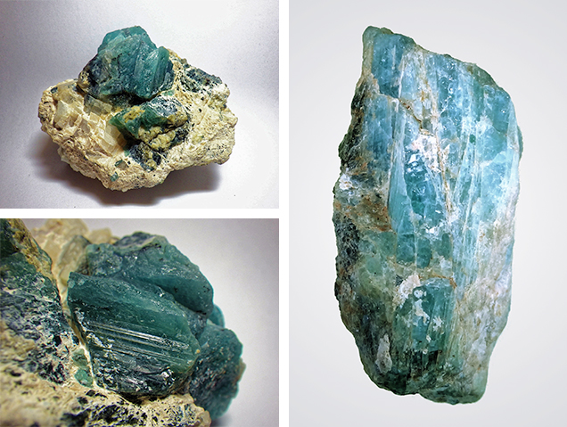
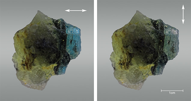
Optical and Physical Properties. The Tranomaro samples consisted of strong bluish green to greenish blue euhedral crystals measuring up to 15 × 7 × 3 cm and weighing 930 g (figure 4). They were typically stubby, and perfect terminations were rare. The samples were generally translucent and only rarely transparent. Although smooth-edged, the crystals showed flat, well-formed faces locally striated following the {100} cleavage. The thickest crystals were opaque, while the thinner transparent crystals were pleochroic, displaying a deep greenish blue color when polarized light was perpendicular to the c-axis (figure 5, left) and a pale yellow to almost colorless hue when the polarized light was parallel to the c-axis (figure 5, right). No fluorescence was observed under either short- or long-wave UV radiation (254 and 365 nm, respectively). The specific gravity of the small pure crystals was established at 3.01 ± 0.02, while that of a rough 39.7 g crystal (figure 4, right), probably influenced by mineral inclusions, was 2.88 ± 0.02. The density of the eye-clean crystals was consistent with Lacroix (1902), which gave a value of 2.99 g/cm3.
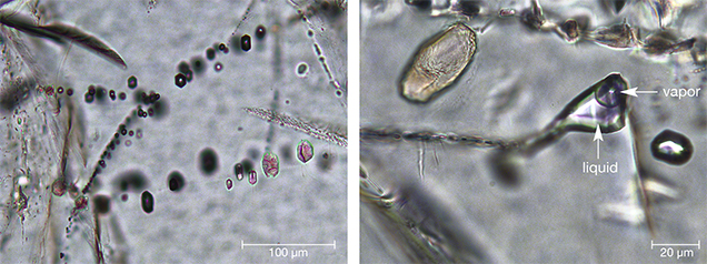
Although the new deposit at Tranomaro was discovered only 80 km northwest of the type locality, there were some slight differences. The recently mined crystals had the same optical properties, but a typically stubby rather than elongated habit. Thin sections observed under the polarizing microscope revealed colorless crystals with a good {100} cleavage. The pleochroism observed visually was not seen in thin section. The crystals were homogenous (without zoning) under both the optical and electron microscopes, and they showed fractures. The minerals were birefringent, with a pink-violet-green color of the second to third orders. Samples from Vein 1 showed lines of birefringent euhedral mineral inclusions 2–40 µm long (figure 6), some with a dipyramidal tetragonal habit consistent with zircon. A few two-phase liquid-vapor fluid inclusions were also present.
Mineral Identification. The XRD patterns of grandidierite from Tranomaro were compared with those obtained on samples from Tolanaro by Stephenson and Moore (1968). Despite the new material’s different habit and specific gravity, no shift was observed in the XRD pattern.
The Raman spectra from the two veins (figure 7) were also similar and revealed several bands that appeared to be characteristic of grandidierite (Maestrati, 1989; Schmetzer et al., 2003; http://rruff.info/R050196). Based on the literature about similar minerals, we propose to relate the spectrum to the mineral structure. Stephenson and Moore (1968) determined that the structure of grandidierite is made up of BO3 triangles; SiO4 tetrahedra; distorted AlO5, MgO5, and FeO5 trigonal bipyramids; and AlO6 octahedra. Vibrations of all functional groups were visible on the Raman spectrum, which could be divided into three regions at 1100–800, 800–400, and 400–100 cm–1. The 1100–800 cm–1 region could be assigned to internal stretching modes of both the planar BO3 group and the SiO4 tetrahedron. The Raman band at 1040 cm–1 could be assigned to the B-O stretching vibration of BO3 units, as with the 1027, 1060, 1086, and 1087 cm–1 bands in other borate minerals (ameghinite, rhodizite, pinakiolite, and takedaite, respectively; see Frost, 2011; Frost and Xi, 2012; Frost et al., 2014a,b). Similarly, the 951 cm–1 band could be assigned to ν1(BO3), since Ross (1974) mentioned a 910–960 cm–1 range corresponding to this mode for six other (Mg,Fe)-borate minerals. The 800–400 cm–1 region can be assigned to bending vibrations of both BO3 and SiO4 units; these modes are commonly reported between 500 and 800 cm–1 for the BO3 group (Galuskina et al., 2008; Maczka et al., 2010; Frost, 2011; Frost et al., 2013; Frost et al., 2014a). Finally, the region below 400 cm–1 probably corresponds to (Al,Mg,Fe)-O vibrations and framework deformation. The ν1 and ν2 modes of the AlO6 octahedron, for example, are known as bands in the 150–200 cm–1 range in kaolinite (Frost et al., 1997).
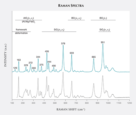
The proposed assignment of Raman peaks to vibrational functional groups must be confirmed by comparison with the mathematical model of the theoretical Raman spectrum for grandidierite.
| TABLE 1. Chemical composition of grandidierite from southern Madagascar by electron microprobe analysis.a | |||||||||
| Oxides (wt.%) |
Vein 1 Sample 1 |
Vein 1 Sample 2 |
Vein 1 Sample 3 |
Vein 1 Sample 4 |
Vein 2 Sample 1 |
Vein 2 Sample 2 |
Vein 2 Sample 3 |
Vein 2 Sample 4 |
Vertriest et al. (2015)b |
| MgO | 14.39 | 14.48 | 14.17 | 14.14 | 14.33 | 14.29 | 14.31 | 14.11 | 17.01 |
| FeOc | 0.61 | 0.51 | 0.45 | 0.53 | 0.53 | 0.47 | 0.53 | 0.56 | 0.30 |
| Al2O3 | 53.02 | 52.91 | 53.12 | 53.29 | 53.61 | 53.25 | 53.23 | 53.23 | 53.89 |
| B2O3 | 12.24 | 9.92 | 9.75 | 11.98 | 8.89 | 10.73 | 9.36 | 9.36 | 12.33 |
| SiO2 | 20.13 | 20.24 | 20.00 | 20.01 | 20.47 | 20.25 | 20.31 | 20.31 | 16.41 |
| Total apfudO9 |
100.39 | 98.05 | 97.49 | 99.95 | 97.83 | 98.99 | 97.75 | 97.75 | 99.95 |
| Mg | 1.03 | 1.07 | 1.05 | 1.02 | 1.07 | 1.04 | 1.06 | 1.06 | 1.23 |
| Fe | 0.02 | 0.02 | 0.02 | 0.02 | 0.02 | 0.02 | 0.02 | 0.02 | 0.01 |
| Al | 3.00 | 3.09 | 3.12 | 3.03 | 3.15 | 3.07 | 3.12 | 3.12 | 3.08 |
| B | 1.01 | 0.85 | 0.84 | 1.00 | 0.77 | 0.91 | 0.80 | 0.80 | 1.03 |
| Si | 0.97 | 1.00 | 1.00 | 0.96 | 1.20 | 0.99 | 1.01 | 1.01 | 0.80 |
| Sum of alkalise |
1.05 | 1.09 | 1.07 | 1.04 | 1.09 | 1.06 | 1.08 | 1.08 | 1.24 |
|
a Ca, K, Mn, Ti, Th, and U are below detection limit (300 ppm for K, Ca, and Ti, and 630 ppm for Mn, Th, and U).
b Laser ablation–inductively coupled plasma–mass spectrometry (LA-ICP-MS) analysis. MnO and Cr2O3 were detected (each 0.01%) but are not included here. c Total iron expressed as FeO. d apfu atoms per formula unit. e Alkalis are Mg and Fe. |
|||||||||
Chemical Composition. Electron microprobe analyses were performed, and the results are listed in table 1. The mineral’s chemical composition was consistent with previously published results and did not vary between the two veins. There were only trace amounts of iron as revealed by the calculated formula (Mg1.02–1.07Fe0.02)Al3.00–3.15(B0.77–1.01O3)(Si0.96–1.02O4)O2. The samples from near Tranomaro had a low iron content of 0.52% FeO, compared to 0.30% FeO in grandidierite recently reported near Tolanaro (Vertriest et al., 2015) and 0.98%–4.27% FeO from elsewhere (Von Knorring et al., 1969; Black, 1970; Seifert and Olesch, 1977; Farges, 2001). As grandidierite is considered the Mg member of the solid solution with ominelite (its Fe analogue), we can also confirm that grandidierite from Madagascar and Johnsburg, New York, constitute the purest grandidierite, with an Fe/(Mg + Fe) ratio below 0.12 (table 2). The studied samples from Tranomaro, along with the material from Johnsburg, have a ratio of 0.04; the grandidierite recently reported near Tolanaro is the purest ever found, with a ratio of 0.02 (Vertriest et al., 2015).
| TABLE 2. Fe/Mg ratio of grandidierite samples worldwide.a | ||
| Locality | Reference | Fe/(Mg + Fe) |
| Tolanaro, Madagascar | Vertriest et al. (2015) | 0.02 |
| Johnsburg, New York | Grew et al. (1991) | 0.04 |
| Tranomaro, Madagascar | This study | 0.04 |
| Tranomaro, Madagascar | This study | 0.05 |
| Tolanaro, Madagascar | Von Knorring et al. (1969) | 0.05 |
| Madagascar | Dzikowski et al. (2007) | 0.08 |
| Vohibola, Madagascar | Seifert and Olesch (1977) | 0.08 |
| Nampoana, Madagascar | Farges (2001) | 0.09 |
| Vohibola, Madagascar | Black (1970) | 0.09 |
| Vohibola, Madagascar | Razakamanana et al. (2010) | 0.10 |
| Sahakondra, Madagascar | Dzikowski et al. (2007) | 0.12 |
| Long Lake, Antarctica | Dzikowski et al. (2007) | 0.14 |
| Zimbabwe | Dzikowski et al. (2007) | 0.14 |
| Faceted gem, Sri Lanka | Schmetzer et al. (2003) | 0.15 |
| Andrahomana, Madagascar | Dzikowski et al. (2007) | 0.15 |
| Karibe area, Zimbabwe | Dzikowski et al. (2007) | 0.17 |
| Almgjotheii, Norway | Dzikowski et al. (2007) | 0.18 |
| Andrahomana, Madagascar | Razakamanana et al. (2010) | 0.27 |
| Sakatelo, Madagascar | Seifert and Olesch (1977) | 0.28 |
| Sakatelo, Madagascar | McKie (1965) | 0.28 |
| Tizi-Ouchen, Algeria | Fabriès et al. (1976) | 0.29 |
| Nampoana, Madagascar | Seifert and Olesch (1977) | 0.33 |
| Nampoana, Madagascar | Farges (2001) | 0.33 |
| Landing Bay, New Zealand | Black (1970) | 0.34 |
| Pedaru, India | Grew (1983) | 0.34 |
| Ampamatoa, Madagascar | Black (1970) | 0.36 |
| Maratakka, Suriname | de Roever and Kieft (1976) | 0.40 |
| McCarthy Pt., Antarctica | Carson et al. (1995) | 0.41 |
| McCarthy Pt., Antarctica | Carson et al. (1995) | 0.41 |
| Westport, Ontario, Canada | Grew (1983) | 0.42 |
| Blanket Bay, New Zealand | Black (1970) | 0.45 |
| Mt. Cimino, Italy | van Bergen (1980) | 0.47 |
| Zambia | Seifert and Olesch (1977) | 0.48 |
| Vestpolltind, Hinnøy, Norway |
Krogh (1975) | 0.48 |
| Ihosy, Madagascar | Nicollet (1990) | 0.49 |
| Mt. Amiata, Italy | van Bergen (1980) | 0.52 |
| Bory Massif, Czech Republic |
Cempírek et al. (2010) | 0.54 |
| Mchinji, Malawi | Haslam (1980) | 0.56 |
| Bory Massif, Czech Republic |
Cempírek et al. (2010) | 0.57 |
| Mchinji, Malawi | Haslam (1980) | 0.59 |
| Ominelite from Japan | Hiroi et al. (2002) | 0.95 |
| Ominelite from Japan | Hiroi et al. (2002) | 0.95 |
| Ominelite from Japan | Hiroi et al. (2002) | 0.96 |
|
a Ominelite specimens were added to compare the Fe/Mg ratio with the grandidierite.
|
||
Compared to the ideal stoichiometric formula for grandidierite, the average Si content was generally consistent, around 1 apfu O9, while locally Al was higher and B was lower (again, see table 1). Because we are confident of the quality of our microprobe analyses, including for B, substitutions might be involved to explain this balance. The substitution of Si by Al or B is well known in other borosilicate minerals, such as some tourmalines, but no substitution of B has hitherto been described in the literature. At this point, with no additional investigations, no further assumption can be made.
Mineral Associations. The two veins were composed mainly of plagioclase, phlogopite, enstatite, and diopside, as identified by XRD. Accessory minerals visible with the unaided eye and identified by SEM-EDS were sapphirine (locally forming gem-quality crystals), chlor-fluorapatite, and dravite.
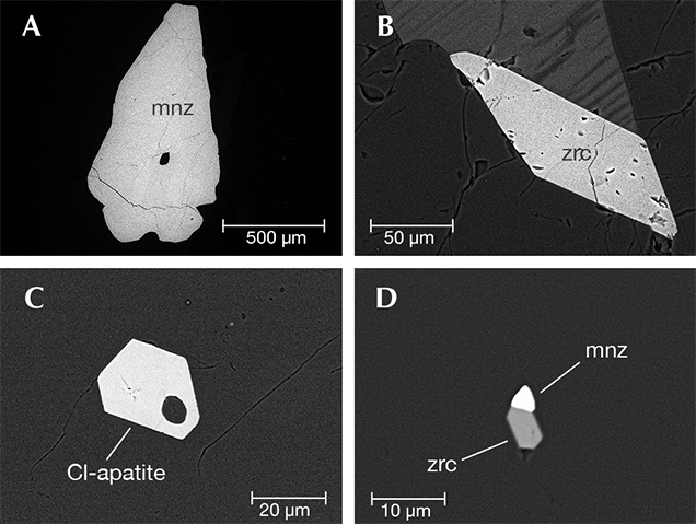
Mineral Inclusions. SEM-EDS observations, confirmed by electron microprobe analyses, also revealed grandidierite crystals hosting Cl-apatite and monazite inclusions of up to a few millimeters long and minute inclusions of zircon (figure 8).
Faceted Stones. Since 2014, a few dozen rough transparent crystals from the Tranomaro deposit have been faceted mainly into round brilliants (again, see figure 1). Some are of gem quality, with transparency and no eye-visible flaws. They display a light blue color in daylight and are trichroic under polarized light (colorless, light blue, and deep greenish blue). Magnification reveals very few flaws. Of these faceted stones, fewer than 10 are above 1 carat. The Tranomaro deposit has produced 800 kg of rough specimens, including about 60 g of eye-clean crystals. The ratio of gem-quality crystals is about 1 in 10,000.
CONCLUSIONS
A new deposit of grandidierite has been discovered in southern Madagascar near Tranomaro, near the now-depleted locality where it was first described. The deposit yields crystals with the same overall properties as those found at the original locality, except that their low iron content and low Fe/(Mg + Fe) ratio make them the purest known grandidierite crystals. The first attempts at faceting provided a few gem-quality stones, including fewer than ten stones above 1 carat. Although modest, this deposit will provide material for museums and collectors in the coming years.


