Andradite in Andradite

Recently we had the opportunity to examine a dramatic iridescent andradite fashioned by Falk Burger (Hard Works, Tucson, Arizona) from a crystal originating from the Tenkawa area of Nara Prefecture in Japan. Known as “rainbow” andradite, this material was previously reported in Gems & Gemology (T. Hainschwang and F. Notari, “The cause of iridescence in rainbow andradite from Nara, Japan,” Winter 2006, pp. 248–258). The specimen was unique for its genesis and optical phenomenon.
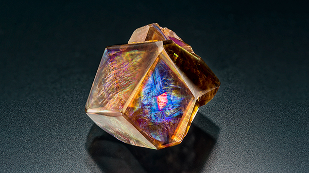
Weighing 16.79 ct and measuring 15.41 × 13.86 × 10.49 mm, the andradite was very large for its species and locality, but size was not what made it special. As shown in figure 1, close examination of one of the polished crystal faces revealed a bright reddish orange “hot spot” in the center, caused by an iridescent inclusion of andradite with a different crystallographic orientation than its host. As seen in figure 2, the inclusion’s different orientation caused the iridescence of the rhomb-shaped “hot spot” to appear and disappear as the light source was passed over the crystal’s surface. To see the iridescence from both the host and inclusion at the same time, two light sources from opposite directions must be used due to the different crystallographic orientation of the host and inclusion. This elusive optical phenomenon made this Japanese andradite crystal extremely interesting for any aspiring inclusionist.


