Natural Faceted Red Rutile
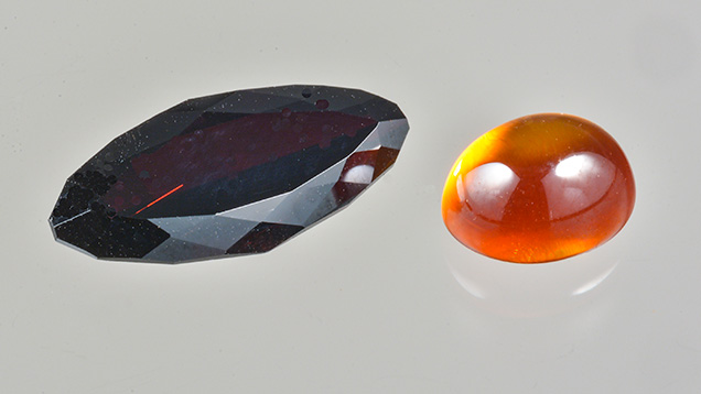
The Carlsbad laboratory recently received a 4.40 ct dark red marquise brilliant (figure 1, left) for examination. Standard gemological testing revealed a refractive index that was over the limit of the RI liquid, as well as a hydrostatic specific gravity of 4.27. The specimen was inert to both long-wave and short-wave UV. Under reflected light, it showed a metallic luster. Raman testing identified the material as rutile. To distinguish it as natural or synthetic rutile, we compared it with a known flame-fusion orange synthetic rutile sample (figure 1, right).

Under magnification, the most distinctive internal characteristic of the dark red specimen was an octagonal blockage with a long growth tube extending to the surface (figure 2). Near the girdle, some unknown rectangular tabular inclusions were also observed, which were not identified by Raman spectroscopy (figure 3). Both internal features indicated the specimen was natural rutile. Straight and angular growth banding was also observed (figure 4). Raman spectroscopy for both the red sample and the orange synthetic revealed major peaks at 610, 446, and 242 cm–1 and minor peaks at 818, 707, and 319 cm–1, confirming their identity as rutile (see the RRUFF database at rruff.info).


Laser ablation–inductively coupled plasma–mass spectrometry (LA-ICP-MS) revealed that the dark red rutile had a composition of 98.2% TiO2 and 1.5% Fe2O3 by weight, with traces of Mg, Al, Si, Ca, Sc, V, Cr, Cu, Zn, Zr, Nb, Cd, In, Sn, Sb, Hf, Ta, W, Pb, and U. The presence of Nb (176.81–296.93 ppmw), Ta (15.13–25.81 ppmw), Hf (0.15–0.40 ppmw), Zr (3.29–4.84 ppmw), Pb (0.01–0.56 ppmw), and U (0.07–0.27 ppmw) suggested a natural origin. The orange synthetic rutile contained 99.9% TiO2, plus traces of Mg, Si, Cr, Co, Cu, and Zn. Traces of iron (1.66 ppmw, 0.0238% Fe2O3 by weight) were also detected. Synthetic rutile boules created by the Verneuil process have an orange color with 0.04% Fe2O3 and a clear reddish color with 0.2% Fe2O3 (C.H. Moore, Jr. and R. Dahlstrom, “Synthetic rutile crystal and method for making same,” U.S. Patent 2792287). The red specimen’s lower chemical purity indicates natural origin.
Visible spectra of both stones were taken. The red bodycolor of the natural rutile matched the transmission window between 650 and 700 nm in the red portion of the spectrum, which was likely caused by iron.
Although rutile is a common inclusion in many minerals, the few crystals that are faceted usually weigh less than 1 ct. This faceted stone was very rare because of its large size. It was the first faceted natural rutile examined by GIA’s Carlsbad laboratory.


