A Large Baroque Tridacna Gigas (Giant Clam) Pearl

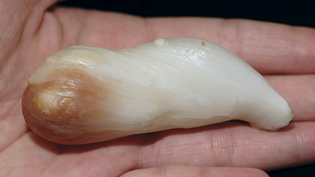
The pearl exhibited an elongated baroque shape with uneven brown and white coloration, lacking the lustrous and shimmering iridescent colors of a nacreous pearl. It weighed 360.59 ct and measured 76.7 × 28.0 × 25.8 mm. We also recorded an SG of 2.88, which fell within the range of other Tridacna pearls examined in our laboratory. Long-wave UV produced a moderate chalky blue reaction. FTIR, Raman, and UV-visible spectra were collected. The Raman spectrum clearly showed that the pearl was composed of aragonite, given the peaks at approximately 142, 199, 701, and 1082 cm–1. FTIR reflectance spectra also revealed calcium carbonate in the form of aragonite, with peaks at approximately 873 and 1483 cm–1.

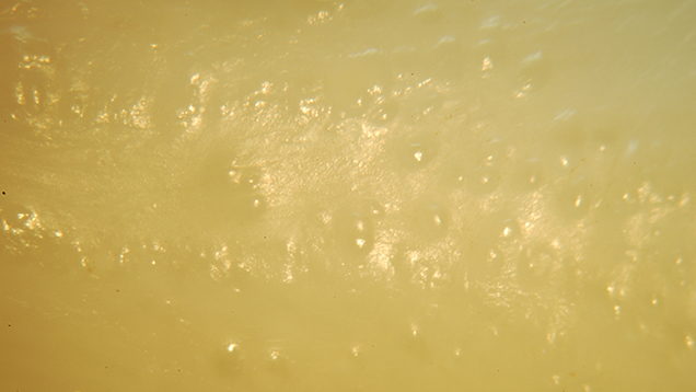
Figure 2. The natural Tridacna pearl showed a characteristic sugary surface texture. Photo by Lai Tai-An Gem Lab; magnified 60×.
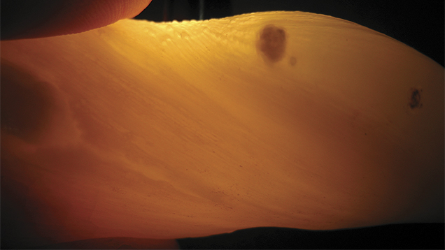
While microradiography is usually applied to the identification of pearls, it is considered especially beneficial when various types of nacreous pearls need to be separated from one another. It is usually less helpful with non-nacreous or non-porcelaneous pearls such as this one, since many reveal little in the way of helpful structure. Observed through the loupe and microscope, the pearl showed the grainy or sugary surface texture (figure 2) typical of some natural Tridacna pearls. This example was noteworthy for its size and interesting coloration, and its pronounced banding when viewed with transmitted light (figure 3), a feature that is often considered indicative of shell fashioned into imitation pearls. This Tridacna pearl was clearly no imitation. 
Figure 3. The Tridacna pearl exhibited pronounced banding when viewed with transmitted light. Photo by Lai Tai-An Gem Lab.


