Pseudo-Synthetic Growth Structure Observed in Natural Diamond

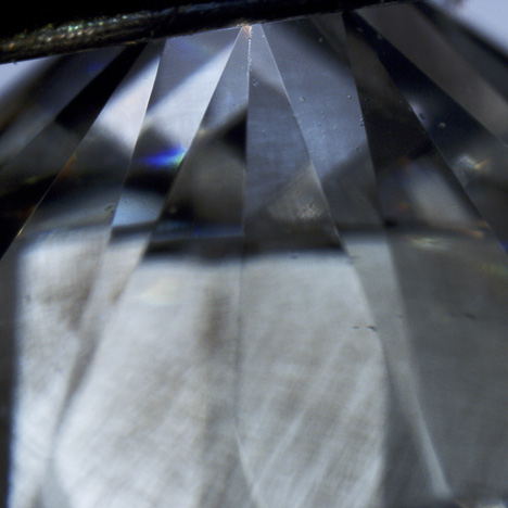
Recently submitted to the GIA laboratory in Israel was a near-colorless 0.70 ct round brilliant (figure 1). Found to be a type IIa diamond with no detectable nitrogen impurity, it was examined further for possible treatments and to verify the origin of color.

Figure 2. DiamondView imaging of the pavilion facets revealed a subtle growth structure. The inset shows the growth structure of a type IIb synthetic diamond. Photo by Wuyi Wang.
In the short-wave UV radiation of the DiamondView, a subtle cuboctahedral growth structure (figure 2) was observed on the pavilion facets. This type of structure usually indicates an HPHT-grown synthetic diamond.The diamond possessed D color and high clarity, with no internal inclusions to help indicate whether it was in fact natural. Shallow surface-reaching fractures were observed, and a few extra facets close to the pavilion contained these natural-looking fractures.
Microscopic observation with cross-polarized light showed relatively strong tatami strain pattern (figure 3), a feature indicative of natural growth. Further examination at higher magnification revealed small polygonal dislocation networks on the pavilion. These provided conclusive evidence that the stone was a natural diamond crystal (figure 4).

Figure 3. A tatami strain pattern, observed under crossed-polarized light, indicated
natural growth. Magnified 40×. Photo by Paul Johnson.
natural growth. Magnified 40×. Photo by Paul Johnson.

Figure 4. DiamondView imaging showed a micro-dislocation network on pavilion facets,
conclusive proof of natural crystal growth. Photo by Wuyi Wang.
This stone was a good example of a very rare natural diamond exhibiting synthetic growth characteristics. It exemplifies the challenges posed to gemological laboratories in separating natural from undisclosed gem-quality synthetic diamonds in today’s jewelry market. We concluded that the diamond had a natural color origin.
conclusive proof of natural crystal growth. Photo by Wuyi Wang.


