Assembled and Bead-Cultured Pearls
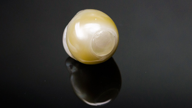
Prior to examination with real-time microradiography, it seemed likely that a shell bead nucleus was hidden from view, as the banded structure of a shell was observed in the translucent area when viewed with transmitted light from a fiber-optic light source. The transparent bonding agent used to join the two parts also contained black impurities (figure 2, left). Obvious gas bubbles were visible with a loupe or gemological microscope. Out of scientific interest, the pearl was also examined with X-ray fluorescence and DiamondView imaging. X-ray fluorescence showed a strong reaction in the damaged and repaired area, as would be expected because of its thinner nacre coverage. DiamondView imaging revealed the banding within the bead, the dark inclusions in the bonding agent, and the bonding agent itself (figure 2, right).

Figure 2. Left: Weak banding was faintly visible beneath the repaired and assembled area when viewed with a strong light source. Field of view 8 mm. Right: The banding was more noticeable in the DiamondView. Photo and DiamondView image by Areeya Manustrong.
Real-time microradiography revealed the bead nucleus as well as an extremely thin nacreous cap that had been used to cover the damaged bead. While X-ray computed microtomography (μ-CT) would not normally be needed to identify such an obviously bead-nucleated pearl, it was used to display the results with even greater clarity (figure 3). This analytical technique clearly showed the broken bead and an area where a new piece of nacre had been applied to repair the pearl. This would explain the mismatching translucent ring.
Figure 3. The broken bead and the nacreous cap applied to conceal the damage are clearly visible in this X-ray computed microtomography (μ-CT) slice.
This study demonstrated how the internal secrets of pearls can be revealed by a variety of techniques.

