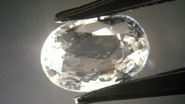
Phosphorescence in Synthetic Sapphire


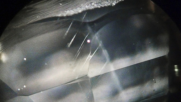
Figure 2. These fine, linear cracks near the girdle resemble long needles. Photomicrograph by Meenakshi Chauhan; magnified 40×.
Magnification showed what appeared to be long needles near the girdle, but closer examination revealed they were long cracks (figure 2). Rather than the Plato lines generally found in light-colored flame-fusion synthetic sapphires, the sample displayed two sharp planes under crossed polarizers. These planes, which had a noticeable separation in between, were visible in twinning lamellae position. Small stress knots near the table surface—but no physical inclusions—were also visible under crossed polarizers (figure 3). Such cracks, which are generally found on the surface of synthetic sapphire, may be due to stress during crystal growth.
Figure 3. These small stress points, visible on the table between crossed polarizers, were confined to a small space. Photomicrograph by Meenakshi Chauhan; magnified 30×.
The sample was inert to long-wave UV radiation but showed a chalky blue fluorescence in short-wave UV, a common feature of synthetic sapphires. It also produced 30 seconds of phosphorescence—the first time this contributor has observed phosphorescence in sapphire, natural or synthetic. Examination in the DiamondView also showed an uncommon blue phosphorescence (figure 4), but it did not show curved growth banding—a fluorescence pattern normally seen in synthetic sapphire grown by the flame-fusion process.
Figure 4. The colorless sample showed blue phosphorescence in the DiamondView, evidence of its synthetic origin. Image by Meenakshi Chauhan.
Identification could only be made on the basis of short-wave UV transmission analysis using the SSEF Diamond Spotter. When the instrument was switched on, short-wave UV radiation transmitted through the sample and the green dot glowed. The specimen’s transparency under the Diamond Spotter’s short-wave emission at 254 nm proved its synthetic origin, as natural sapphire does not transmit short-wave UV below 288 nm (S. Elen and E. Fritsch, “The separation of natural from synthetic colorless sapphire,” Spring 1999 G&G, pp. 30–41, http://dx.doi.org/10.5741/GEMS.35.1.30).Despite the confirmation of synthetic origin, no conclusion could be made regarding the synthesis process used. Consistent with specimens grown by the Czochralski pulling method, there were no curved color bands, inclusions, or Plato lines. Conversely, the specimen’s chalky blue fluorescence and twinning are indicative of flame-fusion growth. These findings present the possibility of either (1) a synthesis process not yet explored by the gemological community or (2) growth by a known process, but with near perfection. While the DiamondView and Diamond Spotter were developed to analyze diamonds, they can also play a vital role in identifying other gemstones.


